. 2011 Jun 3;8(6):639-48.
doi: 10.1016/j.stem.2011.04.003.

Notch-dependent differentiation of adult airway basal stem cells

Affiliations

Notch-dependent differentiation of adult airway basal stem cells

Jason R Rock et al. Cell Stem Cell. .

Abstract

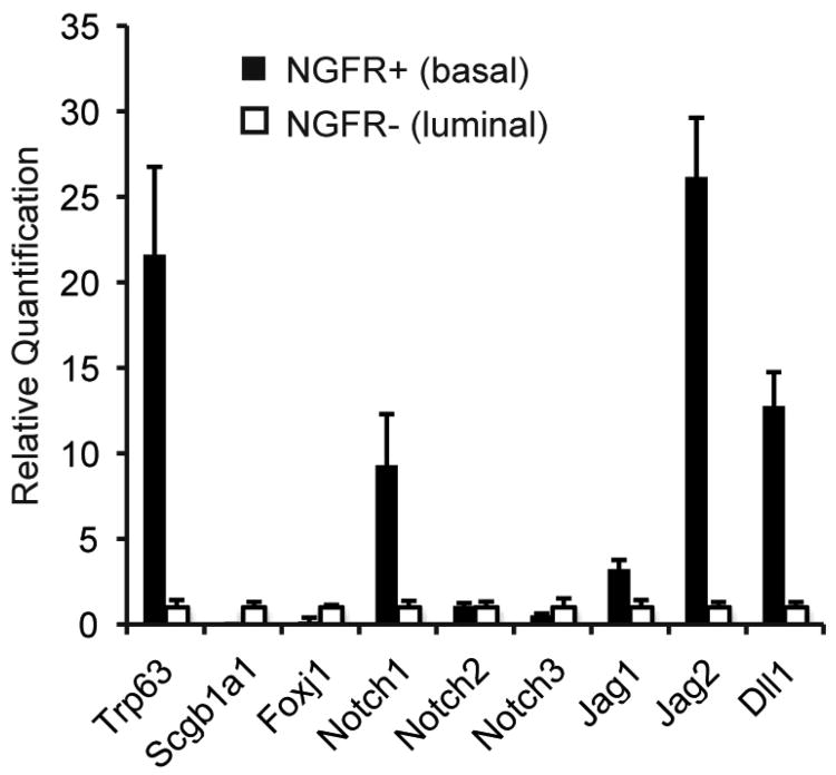
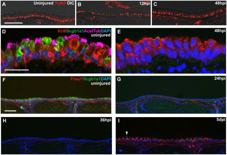
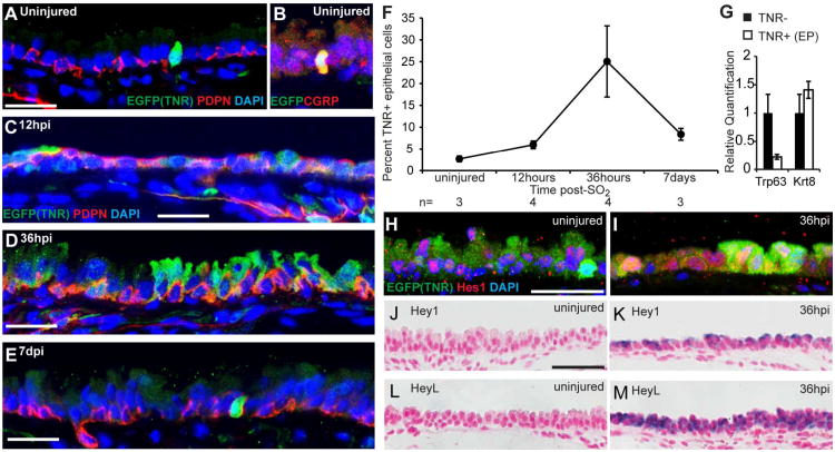
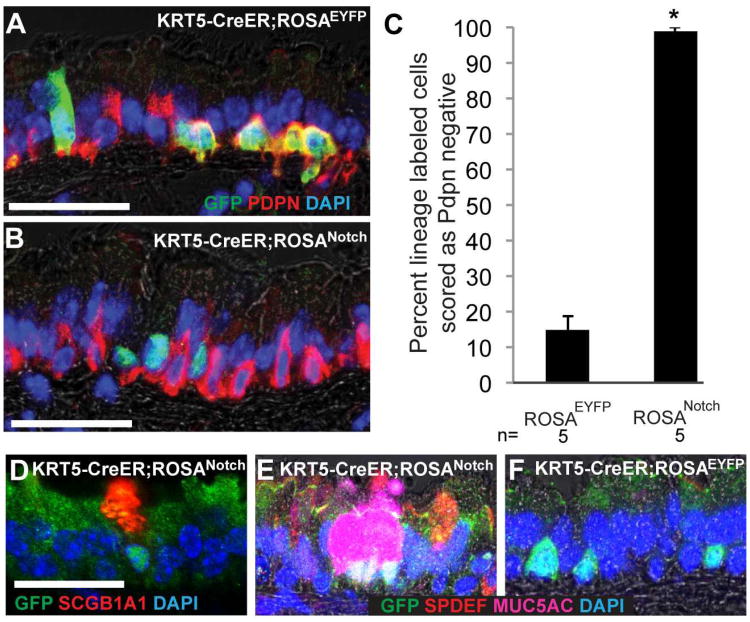
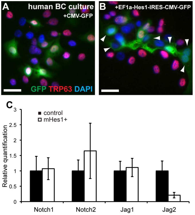
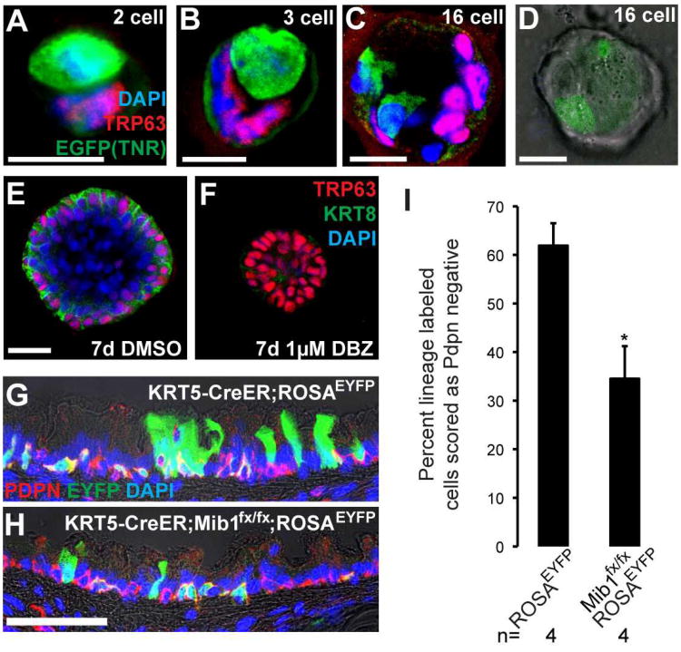
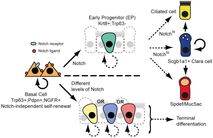
The epithelium lining the airways of the adult human lung is composed of ciliated and secretory cells together with undifferentiated basal cells (BCs). The composition and organization of this epithelium is severely disrupted in many respiratory diseases. However, little is known about the mechanisms controlling airway homeostasis and repair after epithelial damage. Here, we exploit the mouse tracheobronchial epithelium, in which BCs function as resident stem cells, as a genetically tractable model of human small airways. Using a reporter allele we show that the low level of Notch signaling at steady state is greatly enhanced during repair and the generation of luminal progenitors. Loss-of-function experiments show that Notch signaling is required for the differentiation, but not self-renewal, of BCs. Moreover, sustained Notch activation in BCs promotes their luminal differentiation, primarily toward secretory lineages. We also provide evidence that this function of Notch signaling is conserved in BCs from human airways.


Figures

Figure 1
Figure 1. Expression of Notch pathway genes in adult airway epithelium
Expression of Notch pathway components in BCs (black bars) and luminal cells (open bars) assessed by quantitative RT-PCR. y-axis is relative quantification (RQ) normalized to Gapdh and expression of each gene in luminal cells is set to 1 for comparison. Error bars show 95% confidence interval based on triplicate samples. Data shown are representative of three independent experiments.
Figure 2
Figure 2. SO2 inhalation injury model to study mechanisms regulating basal cell behaviors
(A) Control uninjured trachea stained with anti-Trp63 (red, BCs). (B,C) Trp63+ BCs survive SO2 inhalation injury and remain close to the basal lamina 12 hours post-injury (hpi) and during transient stratification 48hpi. Low levels of Trp63 are detected in some suprabasal cells at this time. (D) In the uninjured trachea, ciliated (pink, acetylated tubulin) and Clara (green, Scgb1a1) cells are Krt8+ (red). (E) At 48hpi Krt8+ cells are abundant but negative for markers of ciliated and Clara cells. (F-I) Immunohistochemistry for FoxJ1 (red, ciliated cells) and Scgb1a1 (green, Clara cells) shows these cell types are present in roughly equal proportions in the uninjured trachea. However, neither luminal cell type is observed at (G) 24 or (H) 36hpi. (I) At 5dpi there are abundant FoxJ1+ cells but very few Scgb1a1+ Clara cells (arrowhead). Nuclei were stained with DAPI. Scale bars:A,D 25μm; F 100μm.
Figure 3
Figure 3. Notch signaling is active during epithelial repair
(A-E) Sections of trachea from adult TNR mice stained with anti-GFP to show TNR+ Notch responsive cells (green) and anti-Pdpn (red, BCs) in (A,B) uninjured trachea or (C) 12hpi, (D) 36hpi, and (E) 7dpi. (B) In uninjured tracheas, ∼73% of TNR+ cells are CGRP+ (red, neuroendocrine cells). (F) Percent of epithelial cells scored as TNR+ during repair. Data are means ±SEM and n is the number of tracheas examined at each time. (G) qPCR on cDNA synthesized from TNR- cells (black bars) and TNR+ putative EPs (white bars) obtained by FACS at 36hpi. y-axis is relative quantification (RQ) compared to Gapdh and expression of each gene in TNR- cells (non-EPs) is set to 1 for comparison. Error bars show 95% confidence interval based on triplicate samples. Data are representative of two independent experiments. (H,I) Sections of tracheas from TNR mice stained with anti-Hes1 (red) and anti-GFP. Many luminal and basal cells express Hes1 in the uninjured trachea (H) and at 36hpi (I). In the repairing trachea, the expression of Hes1 is not restricted to GFP-expressing cells. (J-M) RNA in situ hybridization showing expression (purple) of (K) Hey1 and (M) HeyL 36hpi but not in uninjured tracheal epithelium (J,L). Nuclei were stained with DAPI or nuclear fast red. Fluorescent images are single confocal planes. Scale bars:A,C,D,E,H 25μm; J 50μm.
Figure 4
Figure 4. Notch gain-of-function promotes luminal differentiation of mouse airway basal cells in vivo
(A) Adult KRT5-CreER;ROSAEYFP and (B) KRT5-CreER;ROSANotch mice were injected with tamoxifen and sections stained 2 weeks later with anti-GFP (green, lineage label is cytoplasmic for ROSAEYFP and nuclear for ROSANotch) and anti-Pdpn (red, BCs). (C) Percent of lineage labeled cells scored that had differentiated and lost expression of the BC marker Pdpn. Data shown are means ± SEM and n is the number of animals analyzed. *p<0.0001. (D) 81% of cells expressing the Notch1 intracellular domain and nuclear localized GFP stained positively for the Clara cell marker Scgb1a1 and (E) ∼57% were positive for markers of goblet cells including Spdef and Muc5ac. Note that these differentiated cells often appear in pairs or small clusters. (F) Only ∼5% of lineage labeled cells in controls were positive for markers of secretory cells including Scgb1a1, Muc5ac, or Spdef. Scale bars: A,B 40μm; D 30μm.
Figure 5
Figure 5. Notch gain-of-function analysis in human basal cells
(A,B) Primary human BCs were transduced with lentivirus encoding (A) GFP or (B) mHes1 and GFP. After 4 days, 83% of control cells expressing GFP remained TRP63+ while only 39% of BCs expressing mHes1 and GFP were TRP63+. Arrowheads mark GFP+;TRP63- cells. Scale bars: 25μm. (C) qPCR for Notch pathway components was performed on cDNA synthesized from GFP+ cells that were obtained by FACS 5 days after transduction with lentivirus encoding either GFP alone (black bars) or mHes1 and GFP (white bars). y-axis is relative quantification (RQ) compared to Gapdh and expression in control cells is set to 1 for each gene. Error bars show 95% confidence interval based on triplicate samples.
Figure 6
Figure 6. Loss-of-function analysis shows that Notch signaling is required for the luminal differentiation of airway basal cells
(A-D) Tracheospheres grown from single BCs of TNR mice were stained in whole-mount with anti-GFP (green) to show TNR+ Notch responsive cells and anti-Trp63 (red, basal cells) from the 2-16 cell stages. Tracheospheres grown from wild type BCs were cultured with (E) DMSO or (F) gamma secretase inhibitor DBZ (1μM). After 7d, spheres were stained in whole-mount with anti-Trp63 (red, BCs) and anti-Krt8 (green, luminal cells). (G) Adult KRT5-CreER;ROSAEYFP and (H) KRT5-CreER;Mib1fx/fx;ROSAEYFP mice were injected with tamoxifen and exposed to SO2. Two weeks later, after epithelial repair, sections were stained with anti-GFP (green, lineage label), and anti-Pdpn (red, BCs). (I) Percent lineage labeled cells that had differentiated and lost expression of Pdpn. Data shown are means ± SEM and n is the number of anilmals examined. *p<0.05. Nuclei were stained with DAPI and images are single planes from confocal stacks. Scale bars: A-E 10μm; H 50μm.
Figure 7
Figure 7. Models for Notch signaling in the adult pseudostratified airway epithelium
Self-renewal of BCs is Notch-independent, but their luminal differentiation requires canonical Notch signaling. Upper panel: A multipotent early progenitor (EP) has limited capacity for proliferation and gives rise to mature ciliated and secretory cells in response to a second Notch signal. Lower panel: EPs are more lineage-restricted and their fates are specified by Notch input at the time of BC division. In either model, the abundance of EPs and the kinetics of their proliferation and differentiation may differ under steady state conditions versus repair. It is likely that the BC niche is dynamic and comprised of the extracellular matrix, secreted molecules and nearby epithelial, stromal, immune, neuronal, vascular and smooth muscle cells.

Comment in

References

    1. Blanpain C, Lowry WE, Pasolli HA, Fuchs E. Canonical notch signaling functions as a commitment switch in the epidermal lineage. Genes Dev. 2006;20:3022–3035. - PMC - PubMed
    1. Boers JE, Ambergen AW, Thunnissen FB. Number and proliferation of basal and parabasal cells in normal human airway epithelium. Am J Respir Crit Care Med. 1998;157:2000–2006. - PubMed
    1. Borthwick DW, Shahbazian M, Krantz QT, Dorin JR, Randell SH. Evidence for stem-cell niches in the tracheal epithelium. Am J Respir Cell Mol Biol. 2001;24:662–670. - PubMed
    1. Bouras T, Pal B, Vaillant F, Harburg G, Asselin-Labat ML, Oakes SR, Lindeman GJ, Visvader JE. Notch signaling regulates mammary stem cell function and luminal cell-fate commitment. Cell Stem Cell. 2008;3:429–441. - PubMed
    1. Chiba S. Notch signaling in stem cell systems. Stem Cells. 2006;24:2437–2447. - PubMed

Publication types

Substances