. 2011 Jun 6;208(6):1151-62.
doi: 10.1084/jem.20102229. Epub 2011 May 30.

Invariant NKT cells are required for airway inflammation induced by environmental antigens

Affiliations

Invariant NKT cells are required for airway inflammation induced by environmental antigens

Gerhard Wingender et al. J Exp Med. .

Abstract

Invariant NKT cells (iNKT cells) are a unique subset of T lymphocytes that rapidly carry out effector functions. In this study, we report that a majority of sterile house dust extracts (HDEs) tested contained antigens capable of activating mouse and human iNKT cells. HDEs had adjuvant-like properties in an ovalbumin (OVA)-induced asthma model, which were dependent on Vα14i NKT cells, as vaccinated animals deficient for iNKT cells displayed significantly attenuated immune responses and airway inflammation. Furthermore, the administration of HDEs together with OVA mutually augmented the synthesis of cytokines by Vα14i NKT cells and by conventional CD4(+) T cells in the lung, demonstrating a profound immune response synergy for both Th2 cytokines and IL-17A. These data demonstrate that iNKT cell antigens are far more widely dispersed in the environment than previously anticipated. Furthermore, as the antigenic activity in different houses varied greatly, they further suggest that iNKT cell responses to ambient antigens, particular to certain environments, might promote sensitization to conventional respiratory allergens.


Figures
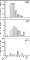
Figure 1.
Figure 1.
HDEs contain antigens for Vα14i NKT cells. (A–C) Microtiter wells coated with CD1d monomers were left untreated (no AG) or were incubated with α-GalCer or the indicated (x axis) HDEs for 18–22 h. Wells were washed and incubated with 5 × 10⁴ of the indicated iNKT cell hybridomas (y axis) for 22–24 h. The Vβ usage of each hybridoma is indicated in the top right of each graph. The IL-2 concentration in the supernatant was determined by ELISA. The depicted values represent the mean of the observed ELISA replicates. Background values have been subtracted. None of the iNKT cell hybridomas responded to HDEs in the absence of CD1d (not depicted). Representative data of at least two experiments are shown. nd, not determined.
Figure 2.
Figure 2.
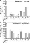
HDEs stimulate human iNKT cell lines. (A) Human PBMCs were left untreated (no AG) or were incubated with α-GalCer or the indicated (x axis) HDEs. After 20 h, cells were washed, and 2 × 10⁵ PBMCs were incubated with 4 × 10⁴ iNKT cells of the human iNKT cell line 59 for 72 h. The GM-CSF concentration in the supernatant was determined by ELISA. Similar results were obtained with the human iNKT cell line 2225 (not depicted). (B) Cell culture plates, coated with CD1d monomers, were left untreated (no AG) or were incubated with α-GalCer or the indicated (x axis) HDEs. After 20 h, plates were washed and incubated with 5 × 10⁴ of the mouse iNKT cell hybridoma DN3A4-1.2 for 22 h. The IL-2 concentration in the supernatant was determined by ELISA. The depicted values represent the mean of the observed ELISA replicates. Background values have been subtracted. Representative data of at least two experiments are shown. Note that samples 1–3 are the same as those tested with DN3A4-1.2 in Fig. 1 A, whereas samples 9–17 were obtained from different houses from those analyzed in Fig. 1.
Figure 3.
Figure 3.
HDE-loaded BM-DCs stimulate mouse iNKT cells in vivo. DCs were generated with GM-CSF from the BM of MyD88 and Trif double-deficient animals (BM-DCDKO; DC in the graphs). BM-DCDKO were washed and incubated with the indicated substances (50 µl/ml HDE, 150 ng/ml LPS, or 50 ng/ml α-GalCer) for 5 h, and 5 × 10⁵ BM-DCDKO were injected i.v. into C57BL/6 mice. 12–15 h later, splenic iNKT cells were analyzed. (A) Relative proportion of iNKT cells in the spleen. (B and C) Representative expression of CD69 by splenic iNKT cells from single mice (B) and a summary of data from at least three mice per group from a representative experiment (C). The numbers in the histogram denote the geometric mean values of CD69 on iNKT cells. (A–C) Representative data from one of four independent experiments, with three to four mice per group, are shown. (D and E) IFN-γ (D) and IL-4 expression (E) by splenic iNKT cells after challenge with the indicated BM-DCDKO. Background values have been subtracted. The graphs in D and E summarize data from three and two independent experiments, respectively, with two to four mice per group. (F–H) Mice were injected with 200 µg α-CD1d antibody (1B1) together with BM-DCDKO loaded with the indicated antigens. Up-regulation of CD69 on iNKT cells (F) or NK cells (H) and production of the indicated cytokines by iNKT cells (G) are depicted. The difference in cytokines production (G) with and without α-CD1d antibody treatment was statistically significant for all cytokines and BM-DCDKO challenges. Representative data from one of two independent experiments, with three to five mice per group, are shown. Error bars indicate mean ± SEM.
Figure 4.
Figure 4.
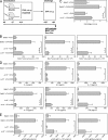
Vα14i NKT cells contribute to the adjuvant effect of HDE during allergen-induced airway inflammation. (A) Experimental outline: BALB/c mice and Jα18-deficient mice on the BALB/c background (Jα18−/−) were sensitized three times, once per week (days 0, 7, and 14) with chicken OVA alone (100 µg/mouse) or together with HDE standard (20 µl/mouse). On days 35 and 41, the mice were challenged with OVA alone (10 µg/mouse) and analyzed 24 h after the last challenge. Abs, antibodies. (B) Total BALF cell count (left) and relative percentages of eosinophils (middle) and neutrophils (right) within BALF from the indicated mice. (C) Histological scores from the lungs of the indicated mice. (D) BLN-derived lymphocytes from the indicated mice were restimulated with 50 µg/ml OVA, and the concentrations of the indicated cytokines in the supernatant were measured by ELISA. (E) OVA-specific antibodies in the sera of the indicated mice including IgE (left), IgG1 (middle), or IgG2a (right). Representative data from one of two independent experiments, with three to four mice per group, are shown. P-values for Jα18−/− + OVA/HDE vs. Jα18−/− + OVA and for Jα18−/− + OVA/HDE vs. BALB/c + OVA/HDE are indicated, where the difference is statistically significant (P, * < 0.05; P, ** < 0.01). Error bars indicate mean ± SEM.
Figure 5.
Figure 5.
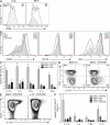
Synergistic activation of lung Vα14i NKT cells and CD4 T cells. (A) BALB/c mice were challenged once with HDE, and the surface expression of CD69 on iNKT cells from lung and BALF was determined 18 h later. The numbers in the histograms denote the geometric mean values of CD69 on iNKT cells. Light gray, closed histogram: BALB/c control; solid line, open histogram: BALB/c challenged with HDE. (B–F) BALB/c mice and Jα18−/− on the BALB/c background were vaccinated three times, once per week (days 0, 7, and 14) with chicken OVA alone (100 µg/mouse), HDE alone, or with OVA together with HDE and analyzed on day 15. Representative data of two or three independent experiments, with two to five mice per group, are shown. (B) Surface expression of CD69, CD122, and CD4 on lung iNKT cells from the indicated groups. The numbers in the histograms denote the geometric mean values of the indicated surface molecules on iNKT cells. (C) Cytokine production of lung iNKT cells ex vivo after the indicated treatments. Lymphocytes were kept 2 h in the presence of degranulation inhibitors to accumulate intracellular cytokines without further stimulation. The graph summarizes data from two or three independent experiments, with three to four mice per group. All cytokine values obtained from the OVA/HDE group were significantly different (P < 0.05) from the values obtained with the other treatments. (D) IL-17A production of lung CD4+ and CD4 iNKT cells from control mice (BALB/c ctr) or from mice challenged with OVA/HDE. (E) CD44 expression and IL-17A production of lung conventional CD4 T cells (CD19 iVα14 CD8α TCR-β+ CD4+) from BALB/c or Jα18−/− mice challenged with OVA/HDE. (F) Cytokine production of lung CD44high conventional CD4 T cells (CD19 iVα14 CD8α TCR-β+ CD4+) ex vivo after the indicated treatments. Lymphocytes were kept 2 h in the presence of degranulation inhibitors to accumulate intracellular cytokines without further stimulation. The graph summarizes data from two or three independent experiments, with three to four mice per group. All cytokine values obtained from the BALB/c + OVA/HDE group were significantly different (P < 0.05) from the values obtained from the Jα18−/− + OVA/HDE group. Error bars indicate mean ± SEM.

References

    1. Adachi O., Kawai T., Takeda K., Matsumoto M., Tsutsui H., Sakagami M., Nakanishi K., Akira S. 1998. Targeted disruption of the MyD88 gene results in loss of IL-1- and IL-18-mediated function. Immunity. 9:143–150 10.1016/S1074-7613(00)80596-8 - DOI - PubMed
    1. Akbari O., Faul J.L., Hoyte E.G., Berry G.J., Wahlström J., Kronenberg M., DeKruyff R.H., Umetsu D.T. 2006. CD4+ invariant T-cell-receptor+ natural killer T cells in bronchial asthma. N. Engl. J. Med. 354:1117–1129 10.1056/NEJMoa053614 - DOI - PubMed
    1. Batzer G., Lam D.P., Paulus P., Boasen J., Ng N., Horner A.A. 2007. Using house dust extracts to understand the immunostimulatory activities of living environments. Immunobiology. 212:491–498 10.1016/j.imbio.2007.03.004 - DOI - PMC - PubMed
    1. Bendelac A., Rivera M.N., Park S.H., Roark J.H. 1997. Mouse CD1-specific NK1 T cells: development, specificity, and function. Annu. Rev. Immunol. 15:535–562 10.1146/annurev.immunol.15.1.535 - DOI - PubMed
    1. Benlagha K., Weiss A., Beavis A., Teyton L., Bendelac A. 2000. In vivo identification of glycolipid antigen–specific T cells using fluorescent CD1d tetramers. J. Exp. Med. 191:1895–1903 10.1084/jem.191.11.1895 - DOI - PMC - PubMed

Publication types