. 2011 Jun 6;208(6):1305-15.
doi: 10.1084/jem.20102419. Epub 2011 May 30.

Hierarchical organization and early hematopoietic specification of the developing HSC lineage in the AGM region

Affiliations

Hierarchical organization and early hematopoietic specification of the developing HSC lineage in the AGM region

Stanislav Rybtsov et al. J Exp Med. .

Abstract

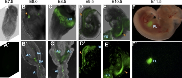
The aorta-gonad-mesonephros region plays an important role in hematopoietic stem cell (HSC) development during mouse embryogenesis. The vascular endothelial cadherin⁺ CD45⁺ (VE-cad⁺CD45⁺) population contains the major type of immature pre-HSCs capable of developing into long-term repopulating definitive HSCs. In this study, we developed a new coaggregation culture system, which supports maturation of a novel population of CD45-negative (VE-cad⁺CD45⁻CD41⁺) pre-HSCs into definitive HSCs. The appearance of these pre-HSCs precedes development of the VE-cad⁺CD45⁺ pre-HSCs (termed here type I and type II pre-HSCs, respectively), thus establishing a hierarchical directionality in the developing HSC lineage. By labeling the luminal surface of the dorsal aorta, we show that both type I and type II pre-HSCs are distributed broadly within the endothelial and subendothelial aortic layers, in contrast to mature definitive HSCs which localize to the aortic endothelial layer. In agreement with expression of CD41 in pre-HSCs, in vivo CD41-Cre-mediated genetic tagging occurs in embryonic pre-HSCs and persists in all lymphomyeloid lineages of the adult animal.


Figures

Figure 1.
Figure 1.
Identification of VE-cad+CD45 pre-HSCs in the AGM region. (A) Experimental design. (B–E) E10.5 (B and C) or E11.5 (D and E) AGM regions were dissociated and sorted on the basis of CD45 and VE-cad expression. Sorted cells were either directly transplanted (B and D) or allowed to mature in coaggregate cultures with OP9 cells for 4 d and then transplanted (C and E). Transplanted doses are 2 (C), 4 (D), and 0.2 ee (E) per recipient. Chimerism (percentage) indicates the proportion of donor-derived cells in the blood of recipient animals. Data are derived from three independent experiments for each type of experiment in C–E. (B) *, see Muller et al. (1994). VC, VE-cad.
Figure 2.
Figure 2.
Quantification of definitive HSCs in coaggregate cultures of the E11.5 AGM region. (A, C, and E) Quantification of definitive HSCs from coaggregate cultures using limiting dilution analysis. (A–D) HSC generation by the VE-cad+CD45 population (A and B) and the VE-cad+CD45+ population (C and D) is shown. 1 ee of the population of interest was aggregated with OP9 cells, and the resulting population was transplanted into each recipient (A, C, and E) at the indicated dose expressed in embryo equivalents (x axis). Each red dot represents one recipient mouse. (B and D) Kinetics of donor-derived blood chimerism upon transplantation of coaggregate cultures of VE-cad+CD45 (B) or VE-cad+CD45+ (D) populations. Each recipient mouse received 1 ee of donor-derived cells. Time after transplantation (months) is indicated. (F) Coaggregates were made with VE-cad+CD45 cells from E11.5 YS, fetal liver, and nonhematopoietic tissues (tail and somites) and transplanted (2 ee/recipient). Control coaggregates of the dorsal aorta were transplanted at 0.2 ee/recipient. Data are derived from three independent experiments for each type of experiments in A–E and two independent experiments for F. VC, VE-cad.
Figure 3.
Figure 3.
In the E11.5 AGM region, VE-cad+CD45 (type I) pre-HSCs mature into definitive HSCs via intermediate VE-cad+CD45+ (type II) pre-HSCs. (A) Time course of appearance of definitive HSCs during 4 d of culture. In each experiment, 1 ee of AGM cells was aggregated with OP9 cells and transplanted into each recipient at each time point. (B) Sorted VE-cad+CD45 cells co-cultured with OP9 cells for 24 h were resorted on the basis of VE-cad (VC) and CD45 expression. All four resorted populations were coaggregated with fresh OP9 cells and cultured for a further 96 h and transplanted separately into irradiated recipients (2 ee/recipient). Numbers of mice repopulated/transplanted are indicated. Data are derived from two independent experiments.
Figure 4.
Figure 4.
In the E11.5 AGM region, CD41 marks type I pre-HSCs and definitive HSCs. (A) Gating strategy for fractionating the VE-cad+CD45 population into CD41high (R1, 46 ± 4/ee), CD41low (R2, 260 ± 18/ee), and CD41 (R3, 2300 ± 110/ee) subsets. R4 includes the VE-cadCD45CD41+ cell population. (B) Recipients were transplanted with sorted populations R1, R2, R3, and R4 after 4-d co-culture with OP9 cells (doses per recipient indicated). (C) AGM region immunostaining for type I pre-HSCs (VE-cad+CD45CD41+). The boxed area within the top left image is shown at high magnification with individual stainings for VE-cad, CD41, or CD45 (top right). Merged images are shown for DAPI and VE-cad (bottom left) and VE-cad, CD41, and CD45 (bottom right). Arrowheads show subluminal localization of these cells. D↔V, dorsoventral orientation. Bar, 10 µm. (D) Gating strategy for subfractionating definitive HSCs on the basis of CD41 expression. (E) VE-cad+CD45+ cells sorted on the basis of CD41 expression (high, R1; low, R2; and negative, R3) from the uncultured AGM region were directly transplanted into irradiated recipients (4 ee/recipient). The CD41low fraction contained HSCs. Data are derived from three independent experiments for all experiments. VC, VE-cad.
Figure 5.
Figure 5.
Lack of endothelial activity in the VE-cad+CD41+ population. (A–D) E10.5 (A and B) and E11.5 (C and D) AGM VE-cad+CD45 populations were purified based on CD41 expression. Error bars show standard deviation of three independent sorting experiments. (A–F) Hematopoietic (A, C, and E) and endothelial (B, D, and F) in vitro assays were set up in 96-well plates using single-cell deposition. (E and F) Hematopoietic (E) and endothelial (F) colonies were stained for CD31 and CD45. DAPI was used to visualize nuclei. Arrows show OP9 cells used as feeders. Bars: (E, left) 200 µm; (E, right) 30 µm; (F and F′) 25 µm. Bar graphs show absolute number of hematopoietic colonies per embryo equivalent (HC/ee; A and C) or endothelial colonies per embryo equivalent (EC/ee; B and D). All data are derived from three independent experiments.
Figure 6.
Figure 6.
Definitive HSCs are localized to the luminal aortic layer, and pre-HSCs are localized to both the endothelial and deeper subendothelial layers. (A and B) Whole-mount preparation of the E11.5 AGM region labeled with OG. (C) Transverse section of OG-labeled E11.5 dorsal aorta (Ao). The intra-aortic cluster is shown in the boxed area. (D and E) Zoomed image of this cluster and aortic endothelium labeled with OG (D) and counter staining with DAPI (E). Bars: (A and B) 700 µm; (C) 50 µm; (D and E) 20 µm. (F) The intensity of fluorescence of cells labeled 1–5 was assessed and plotted as a histogram. Error bars show standard deviations of intensity of fluorescence from five independent confocal images. (G and J) Gating strategy of VE-cad+CD45 (G) and VE-cad+CD45+ (J) cell populations isolated from the E11.5 AGM region on the basis of OG labeling. (H, I, K, and L) Transplantation of freshly isolated cells (H and K) and cells cultured in coaggregate with OP9 stromal cells (I and L). R1, VE-cad+CD45OG cells. R2, VE-cad+CD45OGlow cells. R3, VE-cad+CD45OGhigh cells. R4, VE-cad+CD45+OG cells. R5, VE-cad+CD45+OGlow cells. R6, VE-cad+CD45+OGhigh cells. Dose transplanted was 4 ee/recipient (H and K) and 1 ee/recipient (I and L). Data are derived from two (G–I) or three (J–L) independent experiments. VC, VE-cad.
Figure 7.
Figure 7.
Analysis of CD41-Cre::sGFP embryos. The developing HSC lineage passes through a CD41-positive stage in vivo. (A and A′) E7.5 embryo. (B and B′) E8.0 embryo. GFP+ cells in the YS are indicated by the arrowhead (B) and at the base of the allantois (BA), at the bifurcation of the paired dorsal aorta (Ao), and the allantois (Al) itself (B′). (C and C′) E8.5 embryo. GFP+ cells are within the blood island/belt (BB), allantois, and paired dorsal aorta (C) and within the body of the embryo dissected from the YS (C′). (D and D′) E9.5 embryo. GFP+ cells are in circulation in the dorsal aorta (arrowheads). Bl, blood coming out of the dissected embryo. (E and E′) E10.5 embryo. GFP+ cells in the embryo vasculature. Arrowheads point to the dorsal aorta. (F and F′) E11.5 embryo. GFP+ cells in the liver (FL). A′, D′, E′, and F′ are fluorescent images; A, B, B′, C, C′, D, E, and F are fluorescent images merged with phase-contrast or brightfield images.
Figure 8.
Figure 8.
Level of CD41-Cre–mediated recombination throughout development. (A) GFP expression in the embryonic CD41+ population (gated on CD45 live cells). (B) GFP+VE-cad+CD45 and GFPVE-cad+CD45 populations were purified from E11.5 AGM regions and coaggregated with OP9 cells. After 4 d of co-culture, 0.5 ee of donor-derived cells were transplanted per recipient (each recipient is represented by one bar). Analysis of donor-derived blood cells was performed at 3.5 mo. Data are derived from two experiments. (C) GFP+ and GFP subsets of CD150+CD45+CD48Ter119 cells were sorted from E16.5 fetal livers and transplanted (100 cells/recipient) directly after sorting. Donor-derived blood chimerism was assessed 3.5 mo after transplantation. Each bar represents one recipient. (D) Proportion of GFP+ cells (percentage) in blood and hematopoietic organs of adult CD41-Cre::sGFP animals. (E) Proportion of GFP+ cells (percentage) in different hematopoietic lineages of adult bone marrow. LSK, lineage-negative cKit+Sca1+ cells. LSK150, LinSca1+cKit+CD48CD34low/−CD150+ cells. Each experiment was replicated twice. Error bars show standard deviation (five mice).

References

    1. Bertrand J.Y., Giroux S., Golub R., Klaine M., Jalil A., Boucontet L., Godin I., Cumano A. 2005. Characterization of purified intraembryonic hematopoietic stem cells as a tool to define their site of origin. Proc. Natl. Acad. Sci. USA. 102:134–139 10.1073/pnas.0402270102 - DOI - PMC - PubMed
    1. Bertrand J.Y., Chi N.C., Santoso B., Teng S., Stainier D.Y., Traver D. 2010. Haematopoietic stem cells derive directly from aortic endothelium during development. Nature. 464:108–111 10.1038/nature08738 - DOI - PMC - PubMed
    1. Boisset J.C., van Cappellen W., Andrieu-Soler C., Galjart N., Dzierzak E., Robin C. 2010. In vivo imaging of haematopoietic cells emerging from the mouse aortic endothelium. Nature. 464:116–120 10.1038/nature08764 - DOI - PubMed
    1. Breviario F., Caveda L., Corada M., Martin-Padura I., Navarro P., Golay J., Introna M., Gulino D., Lampugnani M.G., Dejana E. 1995. Functional properties of human vascular endothelial cadherin (7B4/cadherin-5), an endothelium-specific cadherin. Arterioscler. Thromb. Vasc. Biol. 15:1229–1239 - PubMed
    1. Cai Z., de Bruijn M., Ma X., Dortland B., Luteijn T., Downing R.J., Dzierzak E. 2000. Haploinsufficiency of AML1 affects the temporal and spatial generation of hematopoietic stem cells in the mouse embryo. Immunity. 13:423–431 10.1016/S1074-7613(00)00042-X - DOI - PubMed

Publication types