. 2011 Sep 1;20(17):3331-45.
doi: 10.1093/hmg/ddr239. Epub 2011 May 30.

Pathogenity of some limb girdle muscular dystrophy mutations can result from reduced anchorage to myofibrils and altered stability of calpain 3

Affiliations

Pathogenity of some limb girdle muscular dystrophy mutations can result from reduced anchorage to myofibrils and altered stability of calpain 3

Natalia Ermolova et al. Hum Mol Genet. .

Abstract

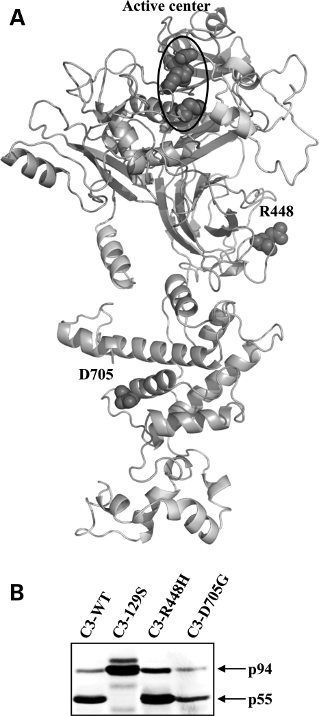
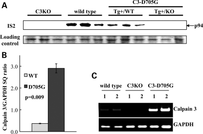
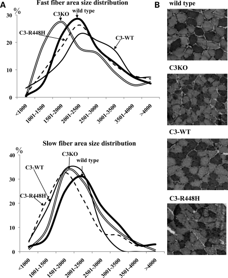
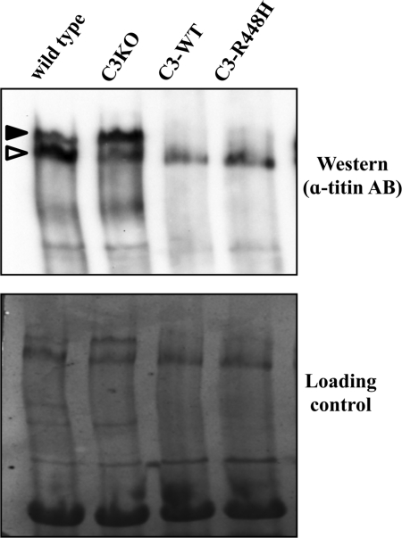
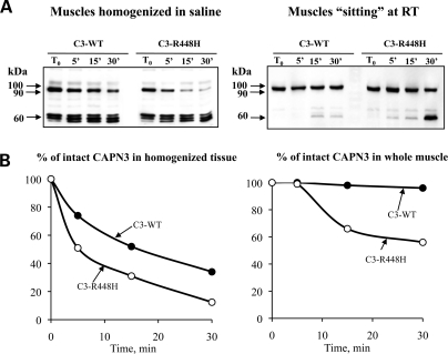
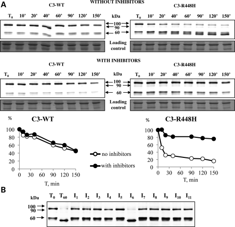
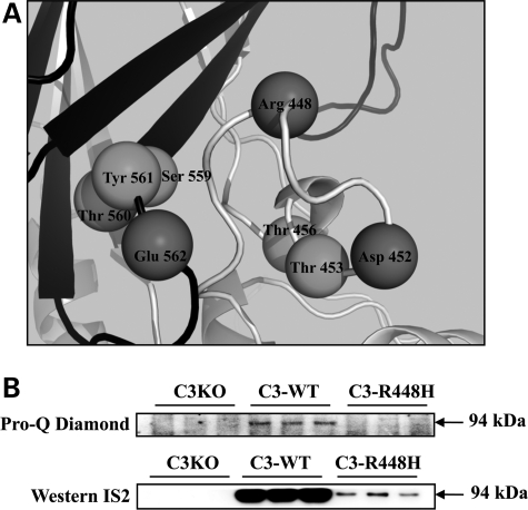
Calpain 3 (CAPN3) is a muscle-specific, calcium-dependent proteinase that is mutated in Limb Girdle Muscle Dystrophy type 2A. Most pathogenic missense mutations in LGMD2A affect CAPN3's proteolytic activity; however, two mutations, D705G and R448H, retain activity but nevertheless cause muscular dystrophy. Previously, we showed that D705G and R448H mutations reduce CAPN3s ability to bind to titin in vitro. In this investigation, we tested the consequence of loss of titin binding in vivo and examined whether this loss can be an underlying pathogenic mechanism in LGMD2A. To address this question, we created transgenic mice that express R448H or D705G in muscles, on wild-type (WT) CAPN3 or knock-out background. Both mutants were readily expressed in insect cells, but when D705G was expressed in skeletal muscle, it was not stable enough to study. Moreover, the D705G mutation had a dominant negative effect on endogenous CAPN3 when expressed on a WT background. The R448H protein was stably expressed in muscles; however, it was more rapidly degraded in muscle extracts compared with WT CAPN3. Increased degradation of R448H was due to non-cysteine, cellular proteases acting on the autolytic sites of CAPN3, rather than autolysis. Fractionation experiments revealed a significant decrease of R448H from the myofibrillar fraction, likely due to the mutant's inability to bind titin. Our data suggest that R448H and D705G mutations affect both CAPN3s anchorage to titin and its stability. These studies reveal a novel mechanism by which mutations that spare enzymatic activity can still lead to calpainopathy.


Figures

Figure 1.
Figure 1.
(A) Structural model of calpain 3 representing the localization of the active center and residues R448 and D705. A monomer of calpain 3 and its four domains is shown. The active center is circled. R448 is located in domain III facing outward; D705 is located at the ‘tip' of EF2 which is one of the calcium-binding EF hands in domain IV. (B) Calpain 3 mutants R448H and D705G can be expressed and demonstrate proteolytic activity in baculoviral cultures. Analysis of WT and mutants (C129S-inactive calpain 3; R448H and D705G) expressed in baculoviral cultures and detected using a calpain 3 specific antibody in western blots (the antibody generated against the IS2 domain is a kind gift from Dr Hiro Sorimachi). C129S migrates as a 94 kDa band as expected for a proteolytically inactive mutant. Both D705G and R448H mutants can be expressed in baculoviral cultures and migrate as a cleaved 55 kDa band as would be expected from autolysis in the IS1 domain.
Figure 2.
Figure 2.
The absence of the D705G mutant protein in muscle extracts is associated with post-transcriptional events. Western blot analysis (A), qPCR (B) and conventional PCR (C) of transgenic skeletal muscle samples from the D705G transgenic mice. (A) Western blot analysis of WT and mutant calpain 3 content in muscle extracts revealed the absence of calpain 3 in C3KO as well as in transgenic positive animals on the C3KO background (Tg+/KO). Note also a dominant negative effect of the D705G transgene on the level of WT CAPN3 in muscle extracts from transgene positive animals on WT background (Tg+/WT). Western blots were done in triplicate for each group of animals. The level of Capn3 mRNA expression is higher in D705G transgene compared with WT as demonstrated by real-time (B) and conventional (C) PCRs suggesting that the loss of D705G is due to post-transcriptional events. GAPDH PCR is used as a cDNA control. Real-time PCR data are shown in graph (B) as the average Starting Quantity (SQ) for Capn3 normalized by SQ for GAPDH. PCR data from two representative samples for each genotype are shown in (C). Vertical bars represent standard error of the mean.
Figure 3.
Figure 3.
The R448H mutant can be stably expressed in transgenic mouse skeletal muscle. Western blot analysis of muscle extracts from transgenic mice demonstrates that the R448H mutant can be detected in vivo. The full length calpain 3 cDNA (C3-WT) was expressed as a transgene to use as a control for the R448H mutant. Characteristic cleavage fragments of 60, 58 and 55 kDa can be seen in extracts from C3-WT and C3-R448H in western blots using the 12A2 antibody.
Figure 4.
Figure 4.
Calpain 3 transgenes rescue the fast fiber atrophy in C3KO, but cannot rescue the slow fiber atrophy. For fiber area size calculations, cross-sections obtained from the mid-belly portion of the soleus muscle (six animals in each group, 10 months old) were stained with monoclonal antibodies to slow myosin heavy chain (A). Fast fiber area distribution (top) and slow fiber area distribution (bottom), %. (B) Slow myosin immunohistochemical staining of the soleus muscle is shown for all genotypes.
Figure 5.
Figure 5.
Mutant and WT transgenes retain in vivo activity against the CAPN3 substrate titin. Western blot (upper panel) analysis of titin gel (lower panel) revealed two titin bands in WT skeletal muscle: upper band (black arrowhead)—intact titin; lower band (white arrowhead)—cleaved titin fragment. Note accumulation of a cleaved titin fragment in WT skeletal muscle; while on the contrary, intact titin accumulated in C3KO. Both C3-WT and C3-R448H transgenes retained proteolytic activity against their in vivo substrate titin.
Figure 6.
Figure 6.
The R448H calpain 3 mutant degrades faster than wild-type calpain 3 in transgenic muscles. (A) Western blot analysis of CAPN3 content at different time points in fresh muscles homogenized in saline (left panel) and in whole muscle allowed to ‘sit' at room temperature for indicated periods of time (right panel). The calpain 3 specific IS2 antibody was used for detection in western blots. (B) Rates of degradation of calpain 3 expressed as a percent of initial concentration of intact protein.
Figure 7.
Figure 7.
The calpain 3 mutant R448H is rapidly degraded by cellular proteases and not through autolytic degradation. (A) Western blot analysis of calpain 3 degradation in the absence and presence of a protease inhibitor cocktail of non-cysteine proteinases (aprotonin, bestatin, pepstatin, AEBSF) shows that degradation of C3-R448H is strongly inhibited, suggesting that the mutation alters protein structure, which makes mutant calpain 3 susceptible to attack from many different cellular proteinases. (B) Inhibition of R448H degradation in homogenized muscles by inhibitors targeting individual protease classes. Inhibitors used: I1-antipain (papain, trypsin, plasmin), I2-aprotonin (serine proteinases), I3-bestatin (amino- and exopeptidases), I4-chymostatin (chymotrypsin), I5-E64 (cysteine proteinases), I6-EDTA (metalloproteinases), I7-leupeptin (serine and cysteine proteinases), I8-pepstatin (aspartic proteinases), I9-AEBSF (serine proteinases), I10-phosphoramidon (metalloproteinases), I11-PMSF (serine proteinases). Both experiments were done with frozen muscle samples and probed with the IS2 antibody.
Figure 8.
Figure 8.
Expression and subcellular localization of GFP-calpain-C129S show co-localization of calpain 3 with the N2 and M line regions of titin. (top panel; versus SHG) (A and B) TPLSM images of an FDB obtained 3 days after transfection with GFP-calpain-C129S. (A) GFP fluorescence, represented in green tones; n is a nucleus and the arrowheads indicate excess fluorescence at the polar regions of the nucleus. (B) Back-scattered second harmonic generation signal, represented arbitrarily by red tones. The asterisks indicate connective tissue. (C) Overlay of images shown in (A) and (B). Graph (D) represents intensity profiles along the rectangles (16 µm) indicated in A (green trace) and B (red trace). The arrowheads indicate the position of the M lines, and the double-headed arrows indicated the positions of the Z lines. (bottom panel; versus di-8-ANEPPS) (A and B) TPLSM images of an FDB obtained 3 days after transfection with GFP-calpain-C129S, and stained with di-8-ANEPPS prior to imaging. (A) GFP fluorescence, represented in green tones. (B) di-8-ANEPPS fluorescence, represented in red tones. (C) Overlay of images shown in A and B. (D) Intensity profiles along the rectangles indicated in A (green trace) and B (red trace). The arrowheads indicate the position of the M-lines, and the double-headed arrows indicated the positions of the Z-lines.
Figure 9.
Figure 9.
Cellular distribution of calpain 3 among various fractions. Fractionation experiments showed calpain 3 present in myofibrillar, cytosolic and membrane fractions with the highest concentration in cytosolic and myofibrillar fractions for both C3-WT and C3-R448H proteins. The amount of calpain 3 in the myofibrillar and membrane fractions was significantly lower when the R448H mutation was present. Western blots and loading controls are shown to the left of the figure. The corresponding densitometric analysis of western blots is shown to the right. Vertical bars represent standard error of the mean.
Figure 10.
Figure 10.
(A) Structural analysis of residues in the vicinity of R448. Shown is the structural model of the area surrounding R448. The potential partners of R448 for salt bridge formation are Asp 452 and Glu 562. Both residues are surrounded by amino acids that could be phosphorylated: Thr 453, Thr 456 near Asp 452 and Ser 559, Thr 560, Tyr 561 near Glu 562. (B) Detection of phosphorylated calpain 3 in the myofibrillar fraction. Pro-Q Diamond phosphoprotein blot stain was used to assess the phosphorylation state of calpain 3 after immunoprecipitation from solubilized myofibrillar fraction. Phosphorylated calpain 3 was not detected in whole extracts subjected to the same procedure. The phosphoprotein was detected more strongly in the myofibrillar fraction of the mice expressing the wild-type transgene. This result might be due to the loss of C3-R448H from the myofibrillar fraction and not necessarily due to differences in the phosphorylation level of calpain 3; thus, quantitative comparisons could not be made in this experiment. Top panel represents the results of staining immunoprecipitated CAPN3 with pro-Q Diamond for the myofibrillar fraction; lower panel represents the same samples detected with CAPN3 specific IS2 antibody.

References

    1. Goll D.E., Thompson V.F., Li H., Wei W., Cong J. The calpain system. Physiol. Rev. 2003;83:731–801. - PubMed
    1. Sorimachi H., Hata S., Ono Y. Expanding members and roles of the calpain superfamily and their genetically modified animals. Exp. Anim. 2010;59:549–566. doi:10.1538/expanim.59.549. - DOI - PubMed
    1. Croall D.E., Ersfeld K. The calpains: modular designs and functional diversity. Genome Biol. 2007;8:218. doi:10.1186/gb-2007-8-6-218. - DOI - PMC - PubMed
    1. Sorimachi H., Suzuki K. The structure of calpain. J. Biochem. 2001;129:653–664. - PubMed
    1. Suzuki K., Sorimachi H., Yoshizawa T., Kinbara K., Ishiura S. Calpain: novel family members, activation, and physiologic function. Biol. Chem. Hoppe Seyler. 1995;376:523–529. doi:10.1515/bchm3.1995.376.9.523. - DOI - PubMed

Publication types

Supplementary concepts