

Chronic oral infection with Porphyromonas gingivalis accelerates atheroma formation by shifting the lipid profile
- PMID: 21625524
- PMCID: PMC3098290
- DOI: 10.1371/journal.pone.0020240
Chronic oral infection with Porphyromonas gingivalis accelerates atheroma formation by shifting the lipid profile
Abstract
Background: Recent studies have suggested that periodontal disease increases the risk of atherothrombotic disease. Atherosclerosis has been characterized as a chronic inflammatory response to cholesterol deposition in the arteries. Although several studies have suggested that certain periodontopathic bacteria accelerate atherogenesis in apolipoprotein E-deficient mice, the mechanistic link between cholesterol accumulation and periodontal infection-induced inflammation is largely unknown.
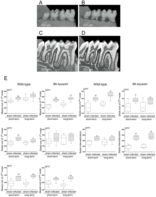
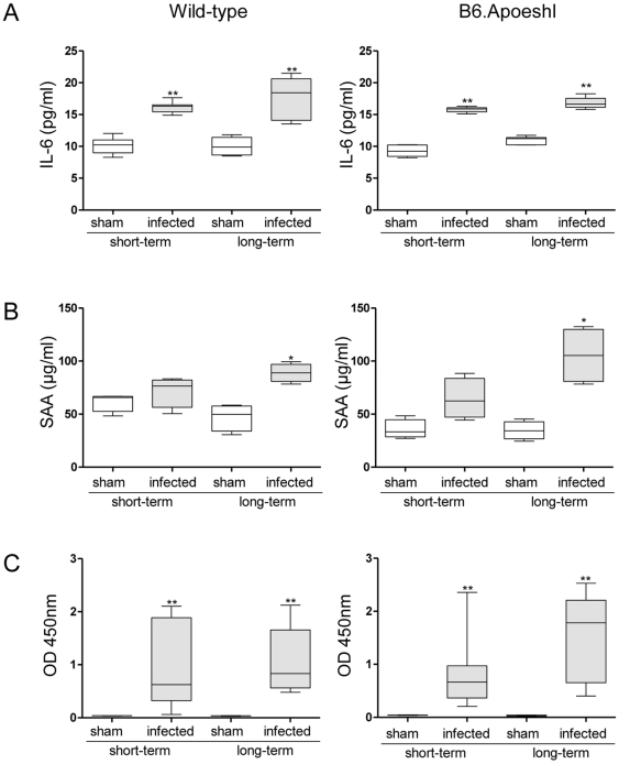
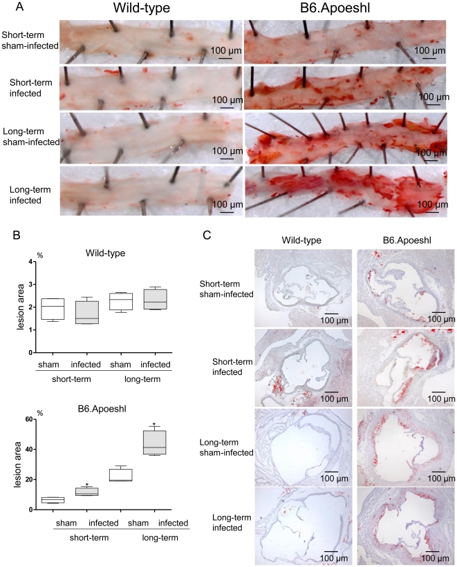
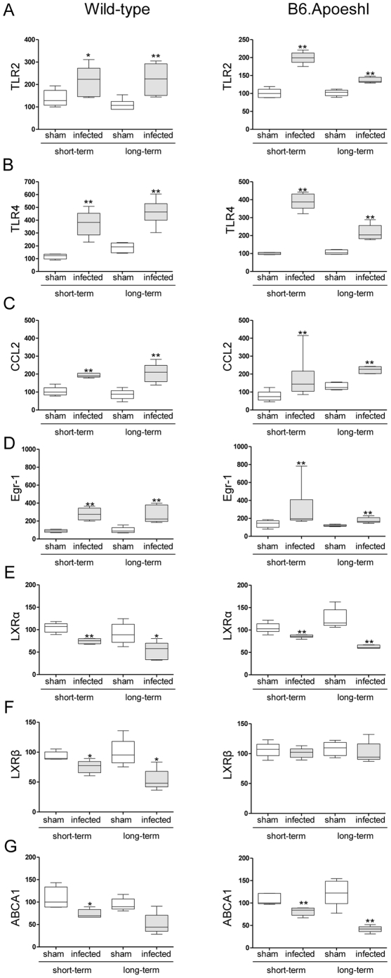
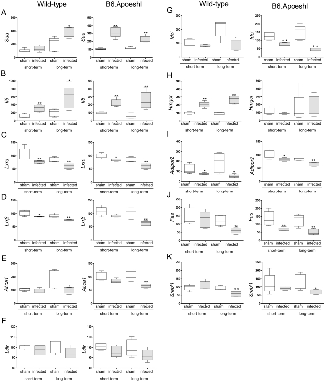
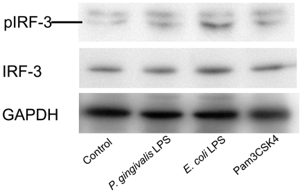
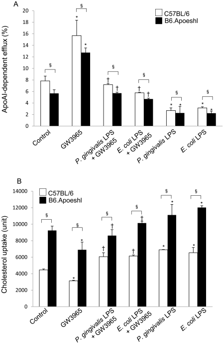
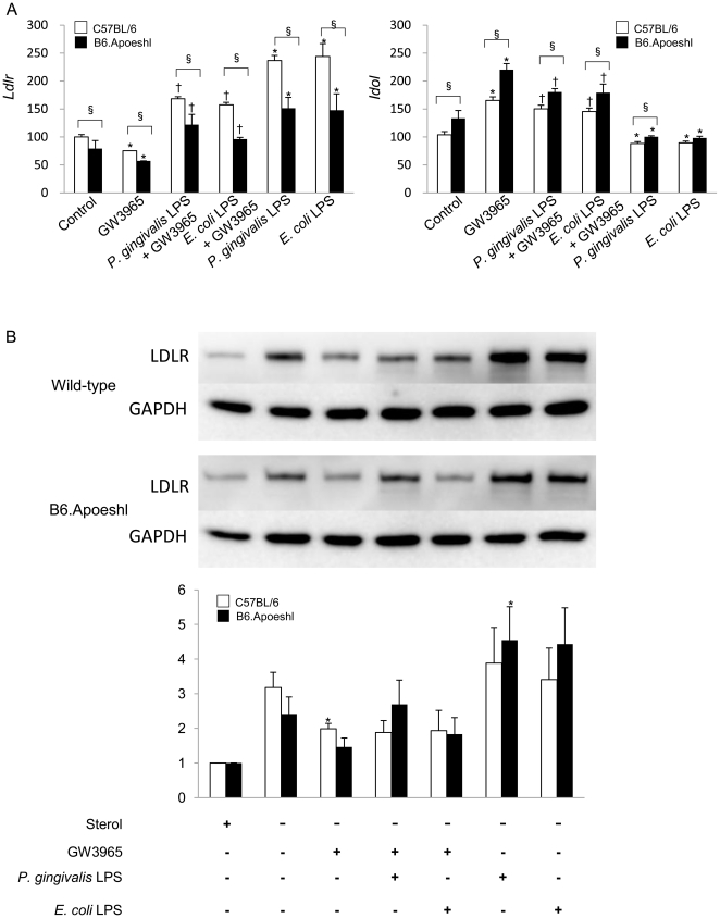
Methodology/principal findings: We orally infected C57BL/6 and C57BL/6.KOR-Apoe(shl) (B6.Apoeshl) mice with Porphyromonas gingivalis, which is a representative periodontopathic bacterium, and evaluated atherogenesis, gene expression in the aorta and liver and systemic inflammatory and lipid profiles in the blood. Furthermore, the effect of lipopolysaccharide (LPS) from P. gingivalis on cholesterol transport and the related gene expression was examined in peritoneal macrophages. Alveolar bone resorption and elevation of systemic inflammatory responses were induced in both strains. Despite early changes in the expression of key genes involved in cholesterol turnover, such as liver X receptor and ATP-binding cassette A1, serum lipid profiles did not change with short-term infection. Long-term infection was associated with a reduction in serum high-density lipoprotein (HDL) cholesterol but not with the development of atherosclerotic lesions in wild-type mice. In B6.Apoeshl mice, long-term infection resulted in the elevation of very low-density lipoprotein (VLDL), LDL and total cholesterols in addition to the reduction of HDL cholesterol. This shift in the lipid profile was concomitant with a significant increase in atherosclerotic lesions. Stimulation with P. gingivalis LPS induced the change of cholesterol transport via targeting the expression of LDL receptor-related genes and resulted in the disturbance of regulatory mechanisms of the cholesterol level in macrophages.
Conclusions/significance: Periodontal infection itself does not cause atherosclerosis, but it accelerates it by inducing systemic inflammation and deteriorating lipid metabolism, particularly when underlying hyperlidemia or susceptibility to hyperlipidemia exists, and it may contribute to the development of coronary heart disease.
Conflict of interest statement
Figures
Similar articles
-
Effect of Porphyromonas gingivalis infection on post-transcriptional regulation of the low-density lipoprotein receptor in mice.Lipids Health Dis. 2012 Sep 19;11:121. doi: 10.1186/1476-511X-11-121. Lipids Health Dis. 2012. PMID: 22992388 Free PMC article.
-
Oral infection with Porphyromonas gingivalis and systemic cytokine profile in C57BL/6.KOR-ApoE shl mice.J Periodontal Res. 2012 Jun;47(3):402-8. doi: 10.1111/j.1600-0765.2011.01441.x. Epub 2011 Nov 20. J Periodontal Res. 2012. PMID: 22097957
-
Porphyromonas gingivalis Infection Accelerates Atherosclerosis Mediated by Oxidative Stress and Inflammatory Responses in ApoE-/- Mice.Clin Lab. 2017 Oct 1;63(10):1627-1637. doi: 10.7754/Clin.Lab.2017.170410. Clin Lab. 2017. PMID: 29035447
-
Porphyromonas gingivalis lipopolysaccharide signaling in gingival fibroblasts-CD14 and Toll-like receptors.Crit Rev Oral Biol Med. 2002;13(2):132-42. doi: 10.1177/154411130201300204. Crit Rev Oral Biol Med. 2002. PMID: 12097356 Review.
-
Pitavastatin: novel effects on lipid parameters.Atheroscler Suppl. 2011 Nov;12(3):277-84. doi: 10.1016/S1567-5688(11)70887-X. Atheroscler Suppl. 2011. PMID: 22152282 Review.
Cited by
-
Cardiovascular Diseases and Periodontitis.Adv Exp Med Biol. 2022;1373:261-280. doi: 10.1007/978-3-030-96881-6_14. Adv Exp Med Biol. 2022. PMID: 35612803 Review.
-
Distinct roles for dietary lipids and Porphyromonas gingivalis infection on atherosclerosis progression and the gut microbiota.Anaerobe. 2017 Jun;45:19-30. doi: 10.1016/j.anaerobe.2017.04.011. Epub 2017 Apr 23. Anaerobe. 2017. PMID: 28442421 Free PMC article.
-
Oral Porphyromonas gingivalis and Fusobacterium nucleatum Abundance in Subjects in Primary and Secondary Cardiovascular Prevention, with or without Heterozygous Familial Hypercholesterolemia.Biomedicines. 2022 Aug 31;10(9):2144. doi: 10.3390/biomedicines10092144. Biomedicines. 2022. PMID: 36140246 Free PMC article.
-
Characterization of Oral Microbiota Following Chemotherapy in Patients With Hematopoietic Malignancies.Integr Cancer Ther. 2023 Jan-Dec;22:15347354231159309. doi: 10.1177/15347354231159309. Integr Cancer Ther. 2023. PMID: 36922730 Free PMC article.
-
Crosstalk between periodontitis and cardiovascular risk.Front Immunol. 2024 Dec 9;15:1469077. doi: 10.3389/fimmu.2024.1469077. eCollection 2024. Front Immunol. 2024. PMID: 39717783 Free PMC article. Review.
References
-
- Chiu B, Viira E, Tucker W, Fong IW. Chlamydia pneumoniae, cytomegalovirus, and herpes simplex virus in atherosclerosis of carotid artery. Circulation. 1997;96:2144–2148. - PubMed
-
- Bahekar AA, Singh S, Saha S, Molnar J, Arora R. The prevalence and incidence of coronary heart disease is significantly increased in periodontitis: a meta-analysis. Am Heart J. 2007;154:830–837. - PubMed
Publication types
MeSH terms
Substances
LinkOut - more resources
Full Text Sources
Medical
Molecular Biology Databases
Miscellaneous
