. 2011;6(5):e20037.
doi: 10.1371/journal.pone.0020037. Epub 2011 May 19.

Large-scale analysis of acute ethanol exposure in zebrafish development: a critical time window and resilience

Affiliations

Large-scale analysis of acute ethanol exposure in zebrafish development: a critical time window and resilience

Shaukat Ali et al. PLoS One. 2011.

Abstract

Background: In humans, ethanol exposure during pregnancy causes a spectrum of developmental defects (fetal alcohol syndrome or FAS). Individuals vary in phenotypic expression. Zebrafish embryos develop FAS-like features after ethanol exposure. In this study, we ask whether stage-specific effects of ethanol can be identified in the zebrafish, and if so, whether they allow the pinpointing of sensitive developmental mechanisms. We have therefore conducted the first large-scale (>1500 embryos) analysis of acute, stage-specific drug effects on zebrafish development, with a large panel of readouts.

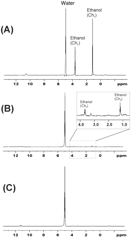
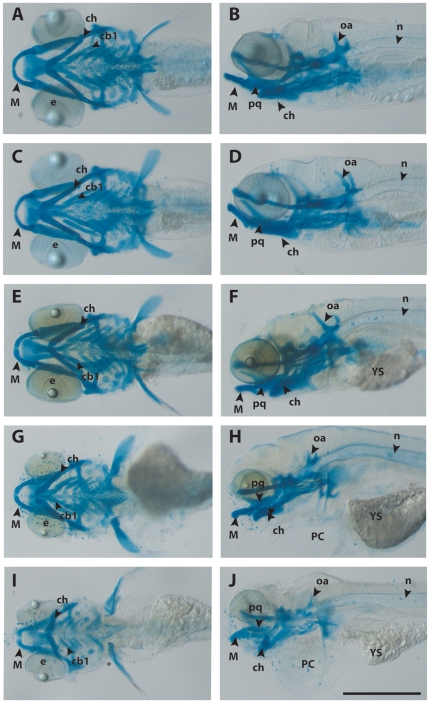
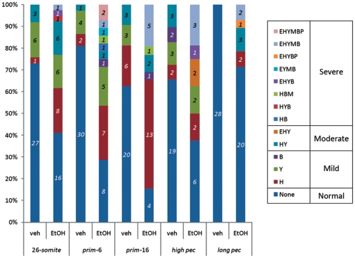
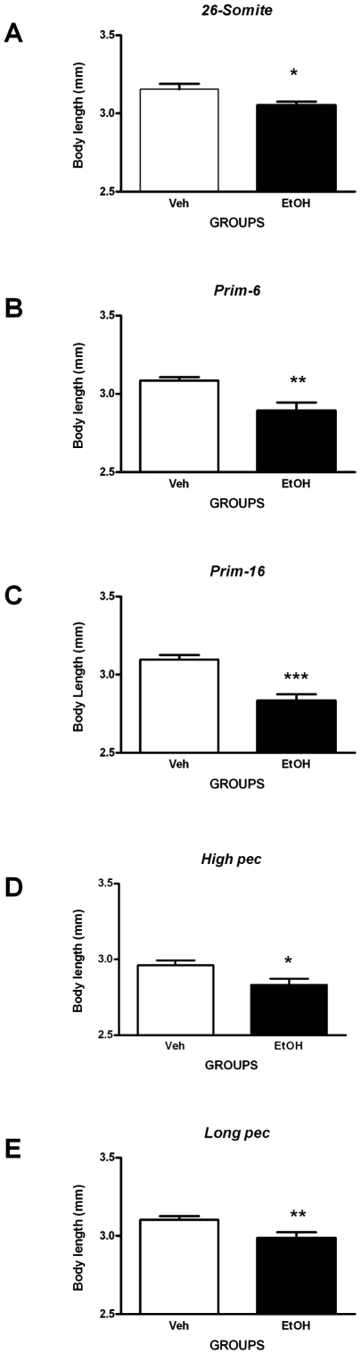
Methodology/principal findings: Zebrafish embryos were raised in 96-well plates. Range-finding indicated that 10% ethanol for 1 h was suitable for an acute exposure regime. High-resolution magic-angle spinning proton magnetic resonance spectroscopy showed that this produced a transient pulse of 0.86% concentration of ethanol in the embryo within the chorion. Survivors at 5 days postfertilisation were analysed. Phenotypes ranged from normal (resilient) to severely malformed. Ethanol exposure at early stages caused high mortality (≥88%). At later stages of exposure, mortality declined and malformations developed. Pharyngeal arch hypoplasia and behavioral impairment were most common after prim-6 and prim-16 exposure. By contrast, microphthalmia and growth retardation were stage-independent.

Conclusions: Our findings show that some ethanol effects are strongly stage-dependent. The phenotypes mimic key aspects of FAS including craniofacial abnormality, microphthalmia, growth retardation and behavioral impairment. We also identify a critical time window (prim-6 and prim-16) for ethanol sensitivity. Finally, our identification of a wide phenotypic spectrum is reminiscent of human FAS, and may provide a useful model for studying disease resilience.


Conflict of interest statement

Competing Interests: The authors have declared that no competing interests exist.

Figures

Figure 1
Figure 1. Survival with a geometric series of ethanol concentrations (1 h exposure), at various developmental stages.
The ethanol concentrations used were: 0, 2, 4, 8, 16 and 32%, Mortality was recorded at various intervals after exposure (48, 72, 96 and 120 hpf).
Figure 2
Figure 2. Ethanol concentration in treated embryos.
Embryos with chorion were treated with 10% ethanol for 1 h and the HR-MAS 1H MRS spectra recorded. A, without subsequent washing; B, after washing three times with buffer. The inset shows a detail enlarged 30 times with respect to the y-axis; C, after washing three times with buffer and subsequently allowed to grow for another 1 h.
Figure 3
Figure 3. Morphological analysis reveals the degree of severity of malformations.
Zebrafish embryos at 5 dpf stained with Alcian blue to show cartilage of the head and branchial region. The aim of this figure is to show examples of the range of severities of malformation obtained (Table 6). A, C, E, G, I, ventral views; B, D, F, H, J, left lateral views. In all figures, rostral is to the left. All embryos are shown to the same scale, indicated by the scale bar (500 µm in J). All embryos were exposed at prim-16 to either vehicle alone (A, B) or 10% ethanol (CJ). A, B, vehicle only, embryo classified as ‘normal’. C, D, ethanol-treated, embryo classified as ‘normal’. E, F, ethanol-treated embryo classified as ‘mild’. The embryo shows yolk sac oedema. G, H, ethanol-treated embryo classified as ‘moderate’. The embryo shows oedema of the yolk sac and pericardium as well as gross microphthalmia. I, J, ethanol-treated embryo, phenotype classified as ‘severe’. The embryo shows gross microphthalmia, pericardial and yolk sac oedema, and grossly hypoplastic Meckel's and branchial cartilages. Key: cb1, 1st ceratobranchial cartilage; ch, ceratohyal cartilage; e, eye; M, Meckel's cartilage; n, notochord; oa, occipital arches; pc, pericardium and heart; pq, palatoquadrate; ys, yolk sac.
Figure 4
Figure 4. Morphology of melanocytes at 5 dpf in embryos treated with ethanol.
All embryos were fixed, stained with Alcian blue and cleared in glycerol. A, embryo treated at long-pec with vehicle only and having a normal phenotype. Note that the melanocytes on the ventral body (arrows) are contracted and punctuate in appearance (scale bar = 250 µm). B, C, embryos treated at high-pec with ethanol and having severe phenotypes (scale bars = 500 µm); note that the melanocytes on the yolk sac (arrows) have a dispersed morphology; in C, the melanocytes on the dorsal surface of the head are also dispersed and form a pavemented layer (arrowheads).
Figure 5
Figure 5. Quantification of melanocyte phenotype at 5 dpf in embryos treated with ethanol at different stages.
‘Contracted morphology’ indicates that the cell is rounded, and the melanosomes concentrated into a small area (Figure 4 A). ‘Dispersed’ morphology (Figure 4 B,C) indicates that the yolk sac melanocytes were squamate and the melanosomes distributed across a wider area than in Figure 4A. As can be seen in the graph, the dispersed morphology is characteristic of ethanol-treated embryos, and reaches a maximum in embryos treated at prim-16. Italic numbers = N embryos.
Figure 6
Figure 6. Stage-dependent sensitivity of the different anatomical regions.
Note that eye development is sensitive to ethanol exposure at all developmental stages (but most sensitive at prim-16). Meckel's cartilage was particularly sensitive to ethanol exposure at prim-6 and prim-16. The branchial arches were most sensitive to ethanol exposure at prim-6, prim-16, and high pec. In contrast to these stage-specific effects, the presence of oedema (i.e. the ‘heart’ and ‘yolk’ categories) was present at low levels following exposure at all stages. Italic numbers = N embryos.
Figure 7
Figure 7. Clustering of morphological abnormalities per embryo.
Number on bars indicates the number of embryos with a particular combination of defects, or single defect, or no gross defect (normal). Surviving embryos were classified according to their phenotype. Key: Normal, no abnormalities; H), embryos with pericardial oedema only; Y, embryos with yolk sac oedema only; B, embryos with branchial arch abnormalities only; HY, embryos with pericardial and yolk sac oedema only; EHY, embryos with microphthalmia, pericardial and yolk sac oedema only; HB, embryos with pericardial oedema and branchial abnormalities only; HYB, embryos with pericardial oedema, yolk sac oedemas and branchial arch abnormalities only; HBM, embryos with pericardial oedema, branchial arch and Meckel's cartilage malformations only; EHYB, embryos with microphthalmia, pericardial oedema, yolk sac oedema and branchial arch defects only; EYMB, embryos with microphthalmia, yolk sac oedema, Meckel's cartilage and branchial arch defects only; EHYBP, embryos with microphthalmia, pericardial oedema, yolk sac oedema, branchial arch and pectoral fin abnormalities only; EHYMB, embryos with microphthalmia, pericardial oedema, yolk sac oedema, Meckel's cartilage and branchial arch abnormalities only; EHYMBP, embryos with microphthalmia, pericardial oedema, yolk sac oedema, Meckel's cartilage, branchial arch and pectoral fin abnormalities only. Italic numbers = N embryos.
Figure 8
Figure 8. Assessment of microphthalmia-like phenotype.
Ethanol treatment is associated with microphthalmia (assayed by measuring eye size at 5 dpf; see Figure S1 B). The graphs show eye size data (µm) for embryos exposed to an acute pulse of 10% ethanol (or vehicle only), for 1 h, at different developmental stages as follows: A, 26-somites; B, prim-6; C; prim-16; D, high-pec; E, long-pec; stages. Statistical analysis (see methods) shows that ethanol exposure at all of these stages except high pec produced significant reduction in eye size (microphthalmia); this effect appears particularly pronounced after exposure at the prim-6, prim-16 and long pec stages. Each error bar represents ±SEM of N = 37, 37, 32, 29, 27 embryos for vehicle and 39, 28, 26, 16, 28 for ethanol treatment at 26-somite, prim-6, prim-16, high pec, and long pec respectively. Statistical icons: ** = p<0.01, and *** = p<0.001.
Figure 9
Figure 9. Assessment of skeletal growth.
Ethanol treatment can produce growth retardation in zebrafish embryos (assayed by measuring body length at 5 dpf; see Figure S1 A). The graphs show body length data (mm) for embryos exposed to an acute pulse of 10% ethanol (or vehicle only), for 1 h, at different developmental stages as follows: A, 26-somites; B, prim-6; C; prim-16; D, high-pec; E, long-pec. Statistical analysis shows that ethanol exposure at all 5 of these stages produced significant growth retardation; this effect was most striking after exposure at the prim-16 stage. Each error bar represents ±SEM of N = 37, 37, 32, 29, 27 embryos for vehicle and 39, 28, 26, 16, 28 for ethanol treatment at 26-somite, prim-6, prim-16, high pec, and long pec respectively. Statistical icons: * = p<0.05, ** = p<0.01, and *** = p<0.001.
Figure 10
Figure 10. Behavioral performance in the light-dark challenge test.
The total distance moved (A, B, C, D and E) and percentage of time spent swimming at high velocity (F, G, H, I and J) were assessed in 5 dpf larvae exposed to the light-dark challenge test. This shows that ethanol-treated embryos swam significantly less (reduced total distance moved) in the challenge phase (lights off) compared to the vehicle-treated controls only when ethanol exposure occurred at prim-16 but not other stages (C). This finding is paralleled (H) by a significantly reduced ability to maintain swimming velocity at a high speed (>20 mm/sec). Furthermore, general decreases in total distance moved, regardless of the phases, are observed in ethanol-treated embryos at stages prim-6 (B) and long-pec (E), suggesting general hypoactivity. This finding is also accompanied by significant reduction in the ability to maintain swimming at high velocity for larvae treated with ethanol at stage prim-6 (G) but not long-pec (J). Note that stages 26-somite (A) and high-pec (D) appear spared from the impact of ethanol exposure on behavioral outcome. Each error bar represents ±SEM of N = 37, 37, 32, 29, 27 embryos for vehicle and 39, 28, 26, 16, 28 for ethanol treatment at 26-somite, prim-6, prim-16, high pec, and long pec respectively. # depicts differences within treatment group. *depicts differences between treatment groups. Statistical icons: ## = p<0.01, * = p<0.05, and ** = p<0.01.

References

    1. Harwood HJ, Fountain D, Fountain G. Economic cost of alcohol and drug abuse in the United States, 1992: a report. Addiction. 1999;94:631–635. - PubMed
    1. Wattendorf DJ, Muenke M. Fetal alcohol spectrum disorders. Am Fam Physician. 2005;72:279–82, 285. - PubMed
    1. Chudley AE, Kilgour AR, Cranston M, Edwards M. Challenges of diagnosis in fetal alcohol syndrome and fetal alcohol spectrum disorder in the adult. Am J Med Genet C Semin Med Genet. 2007;145C:261–272. - PubMed
    1. Spohr HL, Willms J, Steinhausen HC. Fetal alcohol spectrum disorders in young adulthood. J Pediatr. 2007;150:175–9, 179. - PubMed
    1. Moore ES, Ward RE, Wetherill LF, Rogers JL, utti-Ramo I, et al. Unique facial features distinguish fetal alcohol syndrome patients and controls in diverse ethnic populations. Alcohol Clin Exp Res. 2007;31:1707–1713. - PubMed

Publication types

LinkOut - more resources