. 2011;6(5):e19776.
doi: 10.1371/journal.pone.0019776. Epub 2011 May 19.

The participation of calponin in the cross talk between 20-hydroxyecdysone and juvenile hormone signaling pathways by phosphorylation variation

Affiliations

The participation of calponin in the cross talk between 20-hydroxyecdysone and juvenile hormone signaling pathways by phosphorylation variation

Peng-Cheng Liu et al. PLoS One. 2011.

Abstract

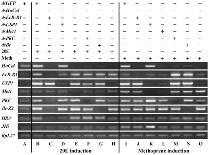
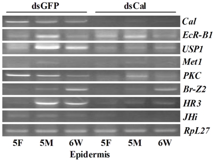
20-hydroxyecdysone (20E) and juvenile hormone (JH) signaling pathways interact to mediate insect development, but the mechanism of this interaction is poorly understood. Here, a calponin homologue domain (Chd) containing protein (HaCal) is reported to play a key role in the cross talk between 20E and JH signaling by varying its phosphorylation. Chd is known as an actin binding domain present in many proteins including some signaling proteins. Using an epidermal cell line (HaEpi), HaCal was found to be up-regulated by either 20E or the JH analog methoprene (JHA). 20E induced rapid phosphorylation of HaCal whereas no phosphorylation occurred with JHA. HaCal could be quickly translocated into the nuclei through 20E or JH signaling but interacted with USP1 only under the mediation of JHA. Knockdown of HaCal by RNAi blocked the 20E inducibility of USP1, PKC and HR3, and also blocked the JHA inducibility of USP1, PKC and JHi. After gene silencing of HaCal by ingestion of dsHaCal expressed by Escherichia coli, the larval development was arrested and the gene expression of USP1, PKC, HR3 and JHi were blocked. These composite data suggest that HaCal plays roles in hormonal signaling by quickly transferring into nucleus to function as a phosphorylated form in the 20E pathway and as a non-phosphorylated form interacting with USP1 in the JH pathway to facilitate 20E or JH signaling cascade, in short, by switching its phosphorylation status to regulate insect development.


Conflict of interest statement

Competing Interests: The authors have declared that no competing interests exist.

Figures

Figure 1
Figure 1. Semi-quantitative RT-PCR analysis of the developmental changes in HaCal mRNA levels.
RpL27 RNA was used as a quantitative control. About 3–20 insects were pooled for each sample at different stages. The values are mean ± S.D. The experiments were repeated at least three times and each was performed in triplicate. Emb: embryos; 1F, 2F, 3F, 4F, 5F: the first to fifth instar feeding larvae; 1 M, 2 M, 3 M, 4 M, 5 M: the first to fifth instar molting larvae; 6–24 h, 6–48 h: sixth instar feeding larvae; 6–72 h, 6–96 h, 6–120 h: sixth instar metamorphic-committed larvae; P0–P9: pupae on day 0 to day 9.
Figure 2
Figure 2. Semi-quantitative RT-PCR analysis of the hormone regulation of HaCal in HaEpi cells.
20E or methoprene was added to the cells to obtain a final concentration of 1 µM. The cells were then cultured for 0.5, 1, 2, 4, and 6 h, respectively, and RNA was extracted for RT-PCR by agarose gel electrophoresis. The values are mean ± S.D. (n = 3). * denoted significant difference (p<0.05), ** denoted significant difference (p<0.01). See also Fig. S1.
Figure 3
Figure 3. Western blot analysis of hormone induction and phosphorylation of HaCal in HaEpi cells.
A, Cultured cells were added with 20E to a final concentration of 1 µM, incubated for 15 and 30 min as well as 1, 3, and 6 h, respectively. Control cells received equal volumes of DMSO. B, Cultured cells were added with methoprene (Meth) to a final concentration of 1 µM. C, The cells were treated with 20E for 30 min, and then the protein was extracted and the partial sample was incubated with λ protein phosphatase (λPP). In addition, protein kinase C specific inhibitor CC (5 µM final concentration) was added to cells shortly before 20E application and incubated for an additional 30 min.
Figure 4
Figure 4. Immunocytochemistry to analyze the effects of 20E (A) and methoprene (B) on HaCal subcellular localization.
Cultured cells were incubated with 20E or methoprene, and then immunostained with anti-HaCal antibody (green color). Nuclei of the cells were stained with DAPI (blue color). HaCal signal was visualized using an Olympus BX51 fluorescence microscope. At least 3 biological replicates were performed and the images are typical ones. a, b, c, and d confirming the subcellular localization of HaCal in the cytoplasm and nucleus by Western blot. Arrow 1 indicates the non-phosphorylated HaCal, and arrow 2 indicates the phosphorylated HaCal. Cy: cytosolic fraction. Nu: nuclear fraction. The yellow bar denotes 20 µm at 40 × magnifications.
Figure 5
Figure 5. Protein interaction between HaCal and USP1.
A, In vitro binding. Recombinant GST-fusion HaCal (GST-Cal) was loaded to the glutathione Sepharose 4B Resin as a bait protein. The recombinant USP1 (His-USP1) was then incubated as a prey. After being washed with 15 mL PBS, the bound proteins were eluted with an elution buffer and separated by 12.5% SDS-PAGE and stained with Commassie blue. B, Recombinant cuticle protein (His-CuP) was used as a negative control for (A). C, In vivo binding of HaCal and USP1. HaEpi cells were incubated with 20E or methoprene. Control cells received equal dilution of DMSO. Total proteins from the treated cells were extracted and precipitated by polyclonal antibody against HaCal through CNBr-activated Sepharose 4B beads. The bound proteins were eluted from the Sepharose beads after extensive washing and subjected to Western blot analysis using antibodies against HaCal and USP1, respectively.
Figure 6
Figure 6. RNAi analysis of the role of HaCal in hormone signaling by RT-PCR in HaEpi cells.
Cells were transfected with dsRNAs of EcR-B1, USP1, Met1, PKC, Br-Z2, and Cal, respectively, and then treated with 20E or methoprene (Meth) for 12 h at a final concentration of 1 µM, respectively. Total RNA was isolated and finally subjected to RT-PCR analysis by agarose gel electrophoresis. An equivalent volume of DMSO was used as solvent control for 20E and methoprene, dsGFP as a non-specific dsRNA control, and RpL27 as a quantitative control. See also Fig. S2.
Figure 7
Figure 7. Effect of HaCal silencing on larval development of H. armigera.
A, Semi-quantitative RT-PCR analysis of transcript levels of HaCal after the larvae ingested bacterially expressed dsHaCal. 5F, 5 M, and 6 W denote 5th instar feeding larvae, 5th instar molting larvae, and 6th instar wandering larvae, respectively. * denotes the significant difference (p<0.05, by student t test). RpL27 was used as a reference. B, Delayed development after knockdown of HaCal. C, Decreased body weight after knockdown of HaCal. D, Postponed body size after knockdown of HaCal. E, Abnormalities after knockdown of HaCal. EcR-B1, USP1, Met1, PKC, Br-Z2, Cal, HR3, and JHi.
Figure 8
Figure 8. Effect of HaCal silencing on the 20E or JH response genes analyzed by RT-PCR.
cDNAs from the epidermis of larvae, in which HaCal was silenced by ingestion of bacterially expressed dsHaCal, were used to check the RNAi effect on these key factors involved in 20E and/or JH signaling pathways. See also Fig. S3.
Figure 9
Figure 9. Chart explaining the function of HaCal in the cross talk between 20E and JH pathways.
A, 20E pathway: 20E signaling leads HaCal protein phosphorylation by PKC via an unknown membrane pathway (1), HaCal is translocated into the nuclei (2), phosphorylated HaCal does not bind with phosphorylated USP1 in 20E pathway (3). EcR binds 20E and USP , and other chaperone protein to form transcription complex, combine the 20E response element initiating the gene transcription , , , , , , . B, JH pathway: Methoprene maintains HaCal non-phosphorylation and translocates HaCal into the nuclei (1), non-phosphorylated HaCal binds with non-phosphorylated USP1 (2). Met binds JH , , and interacts with USP and other chaperone proteins , , and then this complex binds JH response element via Met to initiate JH signaling pathway .

References

    1. Ozlu N, Akten B, Timm W, Haseley N, Steen H, et al. Phosphoproteomics. Wiley Interdiscip Rev Syst Biol Med. 2010;2:255–276. - PubMed
    1. Gilbert LI, Granger NA, Roe RM. The juvenile hormones: historical facts and speculations on future research directions. Insect Biochem Mol Biol. 2000;30:617–644. - PubMed
    1. Riddiford LM, Hiruma K, Zhou X, Nelson CA. Insights into the molecular basis of the hormonal control of molting and metamorphosis from Manduca sexta and Drosophila melanogaster. Insect Biochem Mol Biol. 2003;33:1327–1338. - PubMed
    1. Williams CM. The juvenile hormone II. Its role in the endocrine control of molting, pupation, and adult development in the Cecropia silkworm. Trends Endocrinol Metab. 1961;16:6–11.
    1. Zhou X, Riddiford LM. Broad specifies pupal development and mediates the 'status quo' action of juvenile hormone on the pupal-adult transformation in Drosophila and Manduca. Development. 2002;129:2259–2269. - PubMed

Publication types

MeSH terms