. 2011 May;7(5):e1002052.
doi: 10.1371/journal.ppat.1002052. Epub 2011 May 19.

A gamma interferon independent mechanism of CD4 T cell mediated control of M. tuberculosis infection in vivo

Affiliations

A gamma interferon independent mechanism of CD4 T cell mediated control of M. tuberculosis infection in vivo

Alena M Gallegos et al. PLoS Pathog. 2011 May.

Abstract

CD4 T cell deficiency or defective IFNγ signaling render humans and mice highly susceptible to Mycobacterium tuberculosis (Mtb) infection. The prevailing model is that Th1 CD4 T cells produce IFNγ to activate bactericidal effector mechanisms of infected macrophages. Here we test this model by directly interrogating the effector functions of Th1 CD4 T cells required to control Mtb in vivo. While Th1 CD4 T cells specific for the Mtb antigen ESAT-6 restrict in vivo Mtb growth, this inhibition is independent of IFNγ or TNF and does not require the perforin or FAS effector pathways. Adoptive transfer of Th17 CD4 T cells specific for ESAT-6 partially inhibited Mtb growth while Th2 CD4 T cells were largely ineffective. These results imply a previously unrecognized IFNγ/TNF independent pathway that efficiently controls Mtb and suggest that optimization of this alternative effector function may provide new therapeutic avenues to combat Mtb through vaccination.


Conflict of interest statement

The authors have declared that no competing interests exist.

Figures

Figure 1
Figure 1. Mutation in amino acid 12 of ESAT6 prevents protection by C7 effector cells.
(A) Naïve C7 CD4 T cells were cultured in the presence of irradiated APCs (T cell depleted splenocytes) and either native ESAT6 peptide (aa 1–20) or mutated ESAT6 peptide in which amino acid number 12 was changed from glutamic acid to alanine (E12A). Analysis of tritiated thymidine incorporation reveals C7 CD4 T cells do not respond to ESAT6 (E12A). (B) B6 mice were infected with the indicated strains of Mtb and CFUs determined. Each dot represents data from 4–10 mice. (C) Bacterial numbers from mice that received Th1-skewed C7 effector cells and were subsequently infected with Δesat6+wt or Δesat6+E12A. Bacterial numbers were determined 21 days post infection.
Figure 2
Figure 2. IFN-gamma and iNOS independent control of M. tuberculosis infection.
ESAT-6 specific Th1-skewed CD4 effector cells were transferred into mice which were subsequently aerosol infected with ∼100 CFU of Mtb. Twenty-one days later lungs were harvested and plated to determine bacterial numbers. (A/B) M. tuberculosis numbers in the lungs of WT or IFNγ-deficient mice that either did not receive effector T cells, or received WT or IFNγ-deficient (ko) T cells. (C) Bacterial numbers harvested from either WT, or iNOS ko, mice that either did not receive effector T cells, or received Th1-skewed cells generated from WT ESAT-6 specific CD4 T cells. The data presented represents the combination of 2–3 experiments with 3–5 mice per group. Differences were calculated using unpaired Student's t test.
Figure 3
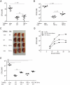
Figure 3. Optimal control of M. tuberculosis growth requires production of IFNγ and TNF by effector T cells.
As in Figure 2, ten million (A–D) or one million (E) Th1-skewed ESAT-6 specific cells were transferred into the indicated hosts that were subsequently infected with Mtb. (A/B) Bacterial numbers in the lungs of WT or TNF deficient (ko) mice that either did not receive effector T cells, or received WT or TNF deficient effector cells. Bacterial numbers were determined 21 days post infection. The data are a combination of 2–3 experiments with 3–4 mice per group. (C) Pictures of lungs from 3 mice from the indicated experimental groups at day 21 post infection. (D) Bacterial numbers at the indicated times harvested from either WT mice that either did not receive cells, or received Th1-skewed WT or IFNγ.TNF dbl deficient cells. Each dot is the average of 7 mice from two independent experiments. Error bars mark SEM. (E) Bacterial numbers from mice receiving either no cells or 1 million C7 effector cells from the indicated donors. Differences were calculated using unpaired Student's t test (A, B, D), or calculated by one way ANOVA (E) ***p<0.0001.
Figure 4
Figure 4. Cytolysis via perforin and FAS are not required for control of M. tuberculosis infection.
(A/B) Th1-skewed ESAT-6 specific cells were transferred into WT mice (expressing CD45.2) that were subsequently transfused i.v. with equal numbers of CD45.1 CFSEhigh, ESAT-6 peptide-pulsed splenocytes, and CFSElow control peptide-pulsed (Listeria lysin O, LLO) splenocytes. (A) In vivo cytotoxicity was assessed 16 hours later by flow cytometry. Dot plots of CFSE and MHC class II levels on CD45.1 splenocytes in recipient mice. The numbers represent frequencies of cells within each quadrant. (B) Cumulative data from two independent experiments from 3–4 mice per group. (C–E) Th1-skewed ESAT-6 specific cells from the indicated backgrounds were transferred into either WT or FAS deficient mice. Bacterial numbers were determined 21 days post infection. Experiments were performed 2 times with 3–5 mice per group. Statistical significance was calculated using the unpaired Student's t test.
Figure 5
Figure 5. Optimal protection to M. tuberculosis requires a Th-1 lineage population, yet is independent of T-bet.
ESAT-6 specific CD4 T cells from the indicated genetic backgrounds were activated for 4 days under Th1, Th2 or Th17 skewing conditions before transfer into the WT mice. Two days after transfer, mice were aerosol infected with ∼100 CFU of Mtb. Twenty-one days later lungs were harvested to determine frequencies and phenotypes of the transferred T cells and lungs were plated to determine bacterial numbers. (A) Frequencies of the transferred populations in the lungs of infected mice, as determined by flow cytometry. (B) Frequencies of IL2, IFNγ, TNF, IL17A, and IL4 producing cells among the transferred effector populations harvested from the lungs. (C) Histogram plots following intracellular staining for the indicated transcription factors. The gate marks the frequencies of RORγt bright cells. (D) Bacterial numbers in the lungs 21 days post infection. *** p<0.0001 calculated by one way ANOVA.

Comment in

References

    1. Cooper AM, Dalton DK, Stewart TA, Griffin JP, Russell DG, et al. Disseminated tuberculosis in interferon gamma gene-disrupted mice. J Exp Med. 1993;178:2243–2247. - PMC - PubMed
    1. Flynn JL, Chan J, Triebold KJ, Dalton DK, Stewart TA, et al. An essential role for interferon gamma in resistance to Mycobacterium tuberculosis infection. J Exp Med. 1993;178:2249–2254. - PMC - PubMed
    1. van de Vosse E, Hoeve MA, Ottenhoff TH. Human genetics of intracellular infectious diseases: molecular and cellular immunity against mycobacteria and salmonellae. Lancet Infect Dis. 2004;4:739–749. - PubMed
    1. Filipe-Santos O, Bustamante J, Chapgier A, Vogt G, de Beaucoudrey L, et al. Inborn errors of IL-12/23- and IFN-gamma-mediated immunity: molecular, cellular, and clinical features. Semin Immunol. 2006;18:347–361. - PubMed
    1. Caruso AM, Serbina N, Klein E, Triebold K, Bloom BR, et al. Mice deficient in CD4 T cells have only transiently diminished levels of IFN-gamma, yet succumb to tuberculosis. J Immunol. 1999;162:5407–5416. - PubMed

Publication types

MeSH terms