Vertical distribution of epibenthic freshwater cyanobacterial Synechococcus spp. strains depends on their ability for photoprotection
- PMID: 21625592
- PMCID: PMC3097228
- DOI: 10.1371/journal.pone.0020134
Vertical distribution of epibenthic freshwater cyanobacterial Synechococcus spp. strains depends on their ability for photoprotection
Abstract
Background: Epibenthic cyanobacteria often grow in environments where the fluctuation of light intensity and quality is extreme and frequent. Different strategies have been developed to cope with this problem depending on the distribution of cyanobacteria in the water column.
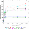
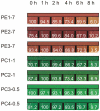
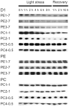
Principal findings: Here we provide an experimental proof that the light intensity plays an important role in the vertical distribution of seven, closely related, epibenthic Synechococcus spp. strains isolated from various water depths from the littoral zone of Lake Constance in Germany and cultivated under laboratory conditions. Pigment analysis revealed that the amount of chlorophyll a and total carotenoids decreased with the time of light stress exposure in three phycoerythrin-rich strains collected from 7.0 m water depth and remained low during the recovery phase. In contrast, a constant level of chlorophyll a and either constant or enhanced levels of carotenoids were assayed in phycocyanin-rich strains collected from 1.0 and 0.5 m water depths. Protein analysis revealed that while the amount of biliproteins remained constant in all strains during light stress and recovery, the amount of D1 protein from photosystem II reaction centre was strongly reduced under light stress conditions in strains from 7.0 m and 1.0 m water depth, but not in strains collected from 0.5 m depth.
Conclusion: Based on these data we propose that light intensity, in addition to light quality, is an important selective force in the vertical distribution of Synechococcus spp. strains, depending on their genetically fixed mechanisms for photoprotection.
Conflict of interest statement
Figures


References
-
- Tandeau de Marsac N. Phycobiliproteins and phycobilisomes: the early observations. Photosynth Res. 2003;76:193–205. - PubMed
-
- MacColl R. Cyanobacterial phycobilisomes. J Struct Biol. 1998;124:311–334. - PubMed
-
- Glazer AN. Light guides. Directional energy transfer in a photosynthetic antenna. J Biol Chem. 1989;264:1–4. - PubMed
-
- Aro EM, Virgin I, Andersson B. Photoinhibition of photosystem II. Inactivation, protein damage and turnover. Biochim Biophys Acta. 1993;1143:113–134. - PubMed
-
- Andersson B, Aro EM. Photodamage and D1 protein turnover in photosystem II. In: Aro EM, Andersson B, editors. Advances in Photosynthesis and Respiration-Regulation of Photosynthesis. Dordrecht: Kluwer Academic Publishers; 2001. pp. 377–393.
Publication types
MeSH terms
Substances
LinkOut - more resources
Full Text Sources
Miscellaneous

