

Experimental alveolitis in rats: microbiological, acute phase response and histometric characterization of delayed alveolar healing
- PMID: 21625744
- PMCID: PMC4234340
- DOI: 10.1590/s1678-77572011000300015
Experimental alveolitis in rats: microbiological, acute phase response and histometric characterization of delayed alveolar healing
Abstract
The pathogenesis of alveolitis is not well known and therefore experimental situations that mimic some features of this disease should be developed.
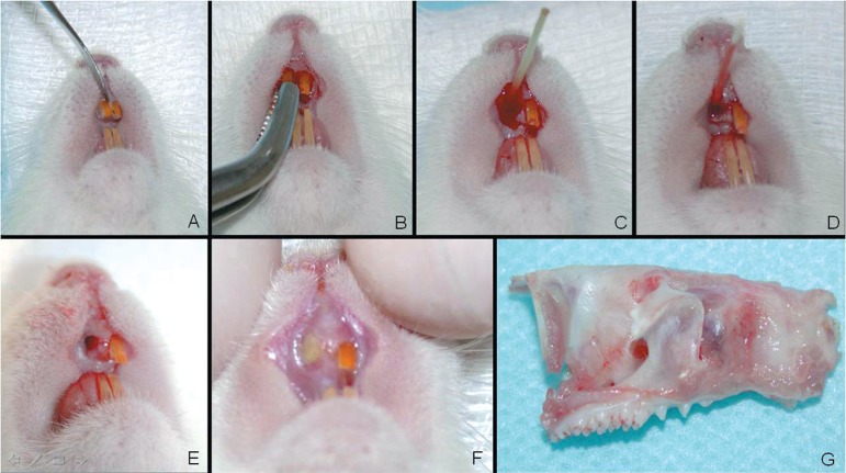
Objective: In this study, the evolution of the experimentally induced infection in rat sockets is characterized, which leads to clinical signs of suppurative alveolitis with remarkable wound healing disturbs.
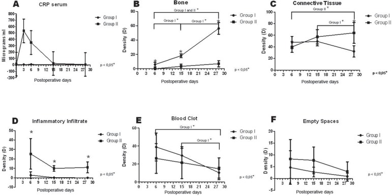
Material and methods: Non-infected (Group I) and experimentally infected sockets in Rattus novergicus (Group II) were histometrically evaluated regarding the kinetics of alveolar healing. In addition, the characterization of the present bacteria in inoculation material and the serum levels of C-reactive protein (CRP) were performed. The detected species were Capnocytophaga ochracea, Fusobacterium nucleatum ss nucleatum, Prevotella melaninogenica, Streptococcus anginosus, Treponema socranskii and Streptococcus sanguis.
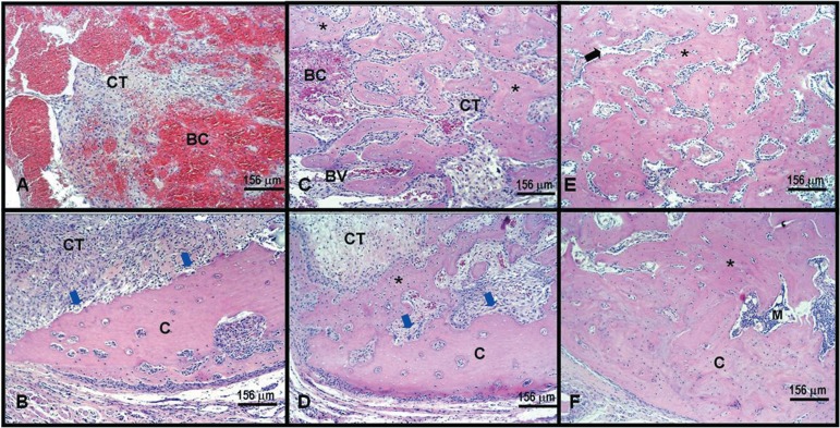
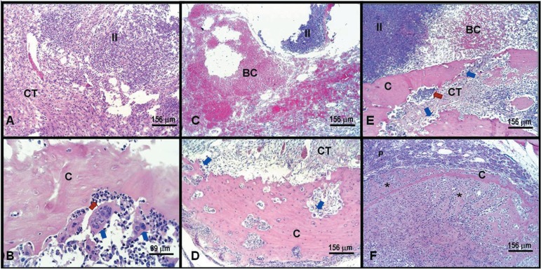
Results: All experimentally infected rats developed suppurative alveolitis, showing higher levels of CRP in comparison to those non-infected ones. Furthermore, infected rats presented a significant delayed wound healing as measured by the histometric analysis (higher persistent polymorphonuclear infiltrate and lower density of newly formed bone).
Conclusion: These findings indicate that rat sockets with experimentally induced infection produced higher levels of serum CRP, showing the potential of disseminated infection and a disturb in the alveolar repair process in an interesting experimental model for alveolitis studies.
Figures
References
-
- Adeyemo WL, Ladeinde AL, Ogunlewe MO. Influence of trans-operative complications on socket healing following dental extractions. J Contemp Dent Pract. 2007;8:52–59. - PubMed
-
- Awang MN. The aetiology of dry socket: a review. Int Dent J. 1989;39:236–240. - PubMed
-
- Baumgartner JC, Khemaleelakul SU, Xia T. Identification of spirochetes (treponemes) in endodontic infections. J Endod. 2003;29:794–797. - PubMed
-
- Birn H. Etiology and pathogenesis of fibrinolytic alveolitis ("dry socket") Int J Oral Surg. 1973;2:211–263.
-
- Cardoso CL, Rodrigues MTV, Ferreira O, Jr, Garlet GP, Carvalho PSP. Clinical concepts of dry socket. J Oral Maxillofac Surg. 2010;68:1922–1932. - PubMed
MeSH terms
Substances
LinkOut - more resources
Full Text Sources
Research Materials
Miscellaneous
