. 2011 Sep 20;44(9):816-27.
doi: 10.1021/ar200037t. Epub 2011 May 31.

Biomedical applications of tetrazine cycloadditions

Affiliations

Biomedical applications of tetrazine cycloadditions

Neal K Devaraj et al. Acc Chem Res. .

Abstract

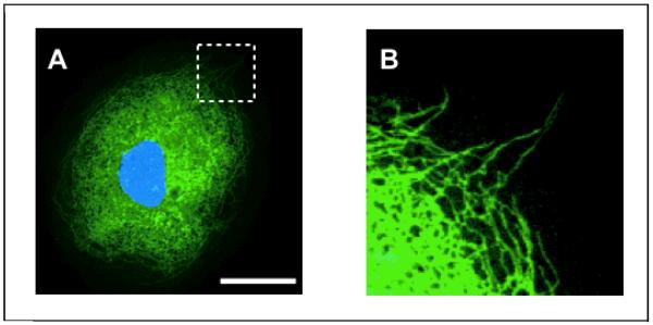
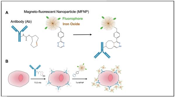
Disease mechanisms are increasingly being resolved at the molecular level. Biomedical success at this scale creates synthetic opportunities for combining specifically designed orthogonal reactions in applications such as imaging, diagnostics, and therapy. For practical reasons, it would be helpful if bioorthogonal coupling reactions proceeded with extremely rapid kinetics (k > 10(3) M(-1) s(-1)) and high specificity. Improving kinetics would minimize both the time and amount of labeling agent required to maintain high coupling yields. In this Account, we discuss our recent efforts to design extremely rapid bioorthogonal coupling reactions between tetrazines and strained alkenes. These selective reactions were first used to covalently couple conjugated tetrazine near-infrared-emitting fluorophores to dienophile-modifed extracellular proteins on living cancer cells. Confocal fluorescence microscopy demonstrated efficient and selective labeling, and control experiments showed minimal background fluorescence. Multistep techniques were optimized to work with nanomolar concentrations of labeling agent over a time scale of minutes: the result was successful real-time imaging of covalent modification. We subsequently discovered fluorogenic probes that increase in fluorescence intensity after the chemical reaction, leading to an improved signal-to-background ratio. Fluorogenic probes were used for intracellular imaging of dienophiles. We further developed strategies to react and image chemotherapeutics, such as trans-cyclooctene taxol analogues, inside living cells. Because the coupling partners are small molecules (<300 Da), they offer unique steric advantages in multistep amplification. We also describe recent success in using tetrazine reactions to label biomarkers on cells with magneto-fluorescent nanoparticles. Two-step protocols that use bioorthogonal chemistry can significantly amplify signals over both one-step labeling procedures as well as two-step procedures that use more sterically hindered biotin-avidin interactions. Nanoparticles can be detected with fluorescence or magnetic resonance techniques. These strategies are now being routinely used on clinical samples for biomarker profiling to predict malignancy and patient outcome. Finally, we discuss recent results with tetrazine reactions used for in vivo molecular imaging applications. Rapid tetrazine cycloadditions allow modular labeling of small molecules with the most commonly used positron emission tomography isotope, (18)F. Additionally, recent work has applied this reaction directly in vivo for the pretargeted imaging of solid tumors. Future work with tetrazine cycloadditions will undoubtedly lead to optimized protocols, improved probes, and additional biomedical applications.


Figures

Figure 1
Figure 1
An example of a tetrazine Diels-Alder cycloaddition. 1,2,4,5 Tetrazines such as 3,6-dimethyl-1,2,4,5-tetrazine can react with dienophiles such as alkenes and alkynes forming formal [4+2] Diels-Alder adducts. These adducts instantly undergo a retro Diels-Alder step, releasing nitrogen. In the case of alkenes, after rearrangement, isomeric dihydropyradazines are typically formed.
Figure 2
Figure 2
A) reaction of benzylamino-tetrazine with trans-cyclooctenol is extremely rapid and leads to dihydropyradazine adducts. B) Live cell multistep labeling of A549 human lung carcinoma cells using tetrazine cycloadditions. Figure reproduced from Ref. [14] with permission from Wiley-VCH.
Figure 3
Figure 3
Confocal microscopy of Cetuximab pretargeted GFP-positive A549 lung cancer cells after tetrazine-fluorophore labeling. A) GFP channel. White scale bar in top left panel denotes 30 microns. B) Red channel: Cetuximab/trans-cyclooctene antibodies have also been directly labeled with AF555 and imaged in the rhodamine channel. C) Near-infrared channel showing the location of bound tetrazine near-infrared probe. D) Merge of GFP, red, and near-infrared channels. Figure reproduced from Ref. [14] with permission from Wiley-VCH.
Figure 4
Figure 4
Real time imaging of tetrazine labeling of pretargeted A549 cells. Cells were exposed to Cetuximab trans-cyclooctene, washed, and imaged in serum (FBS) using the near-IR channel (panel top left). The media was removed and immediately replaced with serum containing 50 nM tetrazine near-infrared probe (top middle panel). Images were taken periodically over 40 minutes. Scale bar in the top left panel denotes 30 microns. Figure reproduced from Ref. [14] with permission from Wiley-VCH.
Figure 5
Figure 5
A) Tetrazine-BODIPY FL reacts rapidly with trans-cyclooctenol via an inverse electron demand Diels-Alder cycloaddition to form isomeric dihydropyrazine products. B) Image comparing the visible fluorescence emission of tetrazine-BODIPY FL (left cuvette) to the corresponding dihydropyrazine products (right cuvette) under excitation from a handheld UV lamp. C) Emission spectra of tetrazine-BODIPY FL (black line) and the corresponding dihydropyrazine products (dashed blue line). Figure reproduced from Ref. [13] with permission from Wiley-VCH.
Figure 6
Figure 6
Confocal imaging of covalently labeled cell-surface bound and intracellular antibodies. A431 cells were exposed to 100 nM of trans-cyclooctene modified anti-EGFR antibody (Cetuximab). Cells were incubated for 1 hour at either 4°C (A) or 37°C (B) and subsequently labeled with 1 μM of a highly charged tetrazine cyanine dye (green) and 1 μM of a lipophilic tetrazine-BODIPY probe (red). Note the intracellular punctate stains representing internalized antibody.
Figure 7
Figure 7
A) Confocal microscopy of a kangaroo rat kidney cell after treatment with 1μM trans-cyclooctene-taxol followed by 1μM tetrazine-BODIPY FL (green). The nucleus is visualized using Hoechst stain (blue). Scale bar: 30 μm. Expansion of the section indicated by the dashed white line (B) reveals intracellular thread-like structures that are clearly stained and visible. Figure reproduced from Ref. [13] with permission from Wiley-VCH.
Figure 8
Figure 8
A) Nanoparticle cycloaddition schematic showing the conjugation chemistry between a trans-cyclooctene modified antibody and tetrazine modified fluorescent iron-oxide nanoparticle. B) Multistep targeting of nanoparticles to specific cellular antigens through use of tetrazine cycloaddition chemistry. Figure reproduced from Ref. [38] with permission from the Nature Publishing Group.
Figure 9
Figure 9
Comparison of different nanoparticle modification techniques for labeling either HER2 (left) or EpCAM (right) biomarkers. Fluorescence intensity of SK-BR-3 and HCT 116 cells labelled with 10 mg ml21 biotin-modified antibody and 100 nM avidin–MFNP was measured using flow cytometry. Biotinylated anti-HER2 and anti-EpCAM antibodies were prepared analogously to the trans-cyclooctene antibodies. Figure reproduced from Ref. [38] with permission from the Nature Publishing Group.
Figure 10
Figure 10
A) Synthesis and structure of 18F-AZD2281 (only one product isomer shown) B) Three-dimensional reconstruction of a tumor-bearing animal injected with 18F-AZD2281 with and without pre-injection of AZD2281 (bladder segmented out for clarity). C) Quantification of uptake through the tumor in hind legs with and without intraperitoneal pre-injection of unlabeled AZD2281. SUV: standardized uptake value. Figure reproduced from Ref. [46] with permission from Wiley-VCH

References

    1. Prescher JA, Bertozzi CR. Chemistry in living systems. Nat Chem Biol. 2005;1:13–21. - PubMed
    1. Baskin JM, et al. Copper-free click chemistry for dynamic in vivo imaging. Proc Natl Acad Sci U S A. 2007;104:16793–16797. - PMC - PubMed
    1. Speers AE, Adam GC, Cravatt BF. Activity-based protein profiling in vivo using a copper(i)-catalyzed azide-alkyne [3 + 2] cycloaddition. J Am Chem Soc. 2003;125:4686–4687. - PubMed
    1. Lewis WG, et al. Click chemistry in situ: acetylcholinesterase as a reaction vessel for the selective assembly of a femtomolar inhibitor from an array of building blocks. Angew. Chem. Int. Ed. 2002;41:1053–1057. - PubMed
    1. Laughlin ST, Baskin JM, Amacher SL, Bertozzi CR. In vivo imaging of membrane-associated glycans in developing zebrafish. Science. 2008;320:664–667. - PMC - PubMed

Publication types