

Review
doi: 10.1021/cr100404w.
Epub 2011 Jun 1.
Mammalian triacylglycerol metabolism: synthesis, lipolysis, and signaling
Affiliations
- PMID: 21627334
- PMCID: PMC3181269
- DOI: 10.1021/cr100404w
Item in Clipboard
Review
Mammalian triacylglycerol metabolism: synthesis, lipolysis, and signaling
Chem Rev.
.
No abstract available
Figures
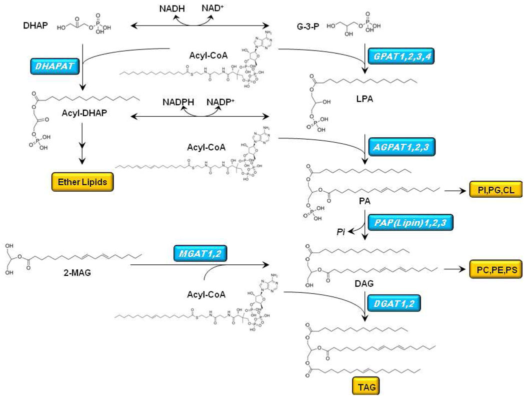
The biosynthesis of TAG begins with the sequential acylation of glycerol-3-phosphate (G-3-P) by sn-1-glycerol-3-phosphate acyltransferase (GPAT) to produce lysophosphatidic acid (LPA) and by acyl-CoA:1-acylglycerol-3-phosphate acyltransferase (AGPAT) to produce phosphatidic acid (PA). G-3-P can also be produced by the oxidation of dihydroxyacetone phosphate (DHAP), and the oxidation of acyl-DHAP produces LPA. PA is hydrolyzed by PA phosphatase (also called lipin) to form diacylglycerol (DAG). A final esterification step by DAG acyltransferase (DGAT) produces triacylglycerol (TAG). PA is also a precursor of CDP-diacylglycerol and the anionic phospholipids phosphatidylglycerol (PG), phosphatidylinositol (PI), and cardiolipin (CL). DAG is a precursor of the phospholipids, phosphatidylcholine (PC), phosphatidylethanolamine (PE), and phosphatidylserine (PS). Acyl-DHAP is the precursor of the ether lipids.
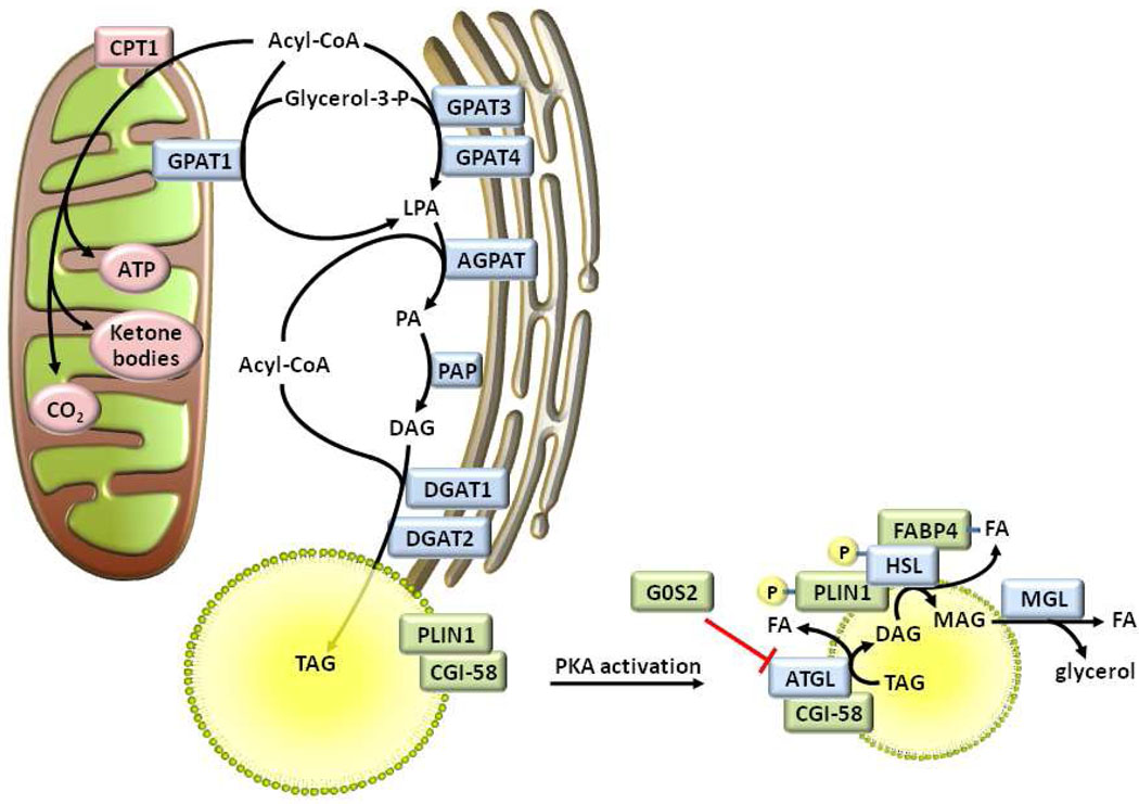
Acyl-CoAs are oxidized in mitochondria after they have been transported into the matrix as acyl-carnitines via carnitine palmitoyltransferase-1 (CPT1). Alternatively, acyl-CoAs may be esterified to glycerol-3-phosphate by a glycerol-3-phosphate acyltransferase (GPAT) isoform, resulting in the production of lysophosphatidic acid (LPA). One or more acyl-CoA:acylglycerol-3-phosphate acyltransferase (AGPAT) isoforms uses a second acyl-CoA to esterify the sn-2 position of LPA to form phosphatidic acid (PA). Phosphatidic acid phosphatase (PAP) dephosphorylates PA to form sn-1,2-diacylglycerol (DAG). Diacylglycerol acyltransferase (DGAT) isoenzymes use a final acyl-CoA to synthesize triacylglycerol (TAG) from DAG. Upon lipolytic stimulation and protein kinase A (PKA) activation, perilipin (PLIN1) is phosphorylated, thereby promoting the release of comparative gene identification-58 (CGI-58) which recruits hormone sensitive lipase (HSL) to the lipid droplet. CGI-58 binds adipose triglyceride lipase (ATGL), facilitates the translocation of ATGL to the lipid droplet, and promotes ATGL’s TAG hydrolytic activity. G0S2 inhibits ATGL, although its interaction with ATGL is not dependent upon PKA activation, as illustrated. PKA also phosphorylates HSL, thereby allowing it to move to the surface of the lipid droplet, and increasing its activity towards DAG. The interaction of HSL with fatty acid binding protein-4 (FABP4) promotes FA efflux from the lipid droplet and alleviates its product inhibition of HSL. Finally, monoacylglycerol lipase (MGL) hydrolyzes monoacylglycerol (MAG), releasing glycerol and a fatty acid (FA).
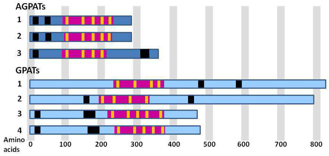
Black regions represent predicted transmembrane domains (PredictProtein; TMHMM; TMpred). Additional transmembrane domains are predicted, but are not shown because they would place sections of the active site region on opposite membrane sides. These predicted domains could interact closely with the membrane. The large rose regions represent the active site region intersected by yellow stripes to indicate the 4 or 5 conserved motifs. Only the topography of GPAT1 has been confirmed experimentally. All active site regions face the cytosol.
References
Publication types
MeSH terms
Substances
Grants and funding
LinkOut - more resources
Full Text Sources
Other Literature Sources
