Review
. 2011 Oct 1;3(10):a004820.
doi: 10.1101/cshperspect.a004820.

Role of lipids in virus replication

Affiliations
Review

Role of lipids in virus replication

Maier Lorizate et al. Cold Spring Harb Perspect Biol. .

Abstract

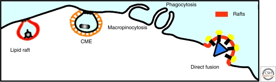
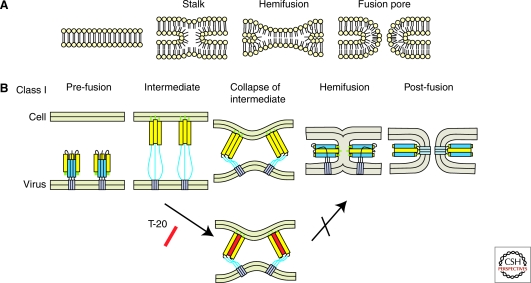
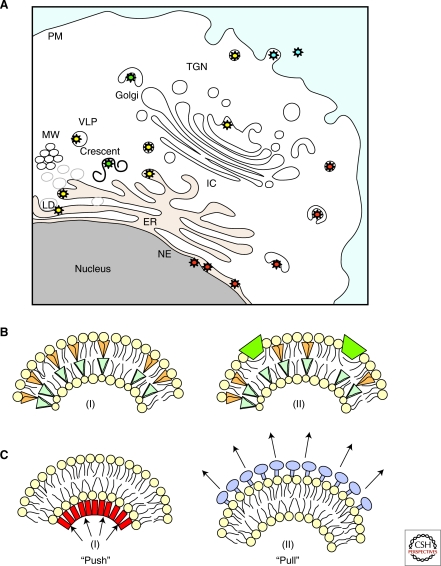
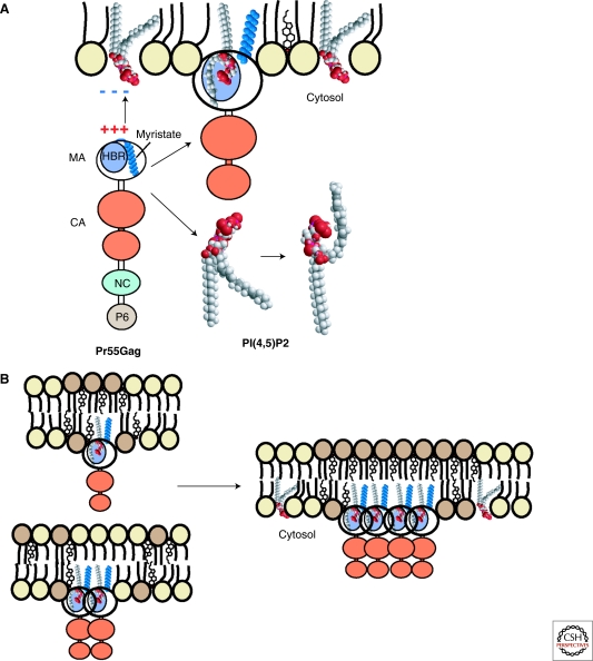
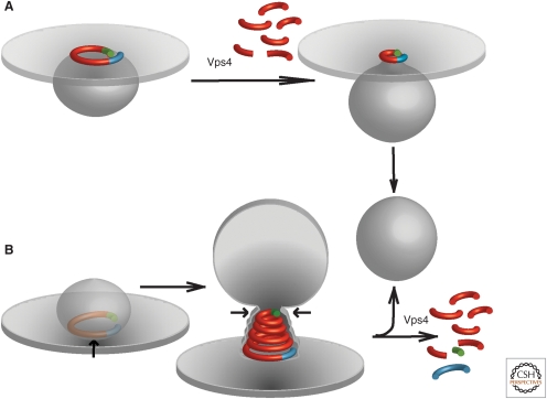
Viruses intricately interact with and modulate cellular membranes at several stages of their replication, but much less is known about the role of viral lipids compared to proteins and nucleic acids. All animal viruses have to cross membranes for cell entry and exit, which occurs by membrane fusion (in enveloped viruses), by transient local disruption of membrane integrity, or by cell lysis. Furthermore, many viruses interact with cellular membrane compartments during their replication and often induce cytoplasmic membrane structures, in which genome replication and assembly occurs. Recent studies revealed details of membrane interaction, membrane bending, fission, and fusion for a number of viruses and unraveled the lipid composition of raft-dependent and -independent viruses. Alterations of membrane lipid composition can block viral release and entry, and certain lipids act as fusion inhibitors, suggesting a potential as antiviral drugs. Here, we review viral interactions with cellular membranes important for virus entry, cytoplasmic genome replication, and virus egress.


Figures

Figure 1.
Figure 1.
Pathways of viral entry. Viruses achieve host cell entry in two principal ways: by direct fusion at the plasma membrane or following an endocytic pathway. The major endocytic pathways operating in mammalian cells that are exploited by viruses are clathrin-mediated endocytosis (CME), lipid raft pathway, clathrin-independent pathways, macropinocytosis, and phagocytosis.
Figure 2.
Figure 2.
Membrane fusion. (A) Stalk mechanisms of lipid bilayer fusion. (B) Fusion models promoted by class I fusion proteins. The lower panel depicts the T-20 mode of action inhibiting transition from the prehairpin structure to six-helix bundle formation by direct binding to the intermediate.
Figure 3.
Figure 3.
Membranes implicated in enveloped virus budding and mechanisms of curvature induction. (A) Virus envelopment can occur at the plasma membrane (particles depicted in blue) or into the lumen of organelles (i.e., ER, LD, Golgi, and TGN) along the secretory pathway (particles depicted in yellow and green). Herpesviruses undergo sequential envelopment, de-envelopment and re-envelopment that take place at the nucleus and TGN (particles depicted in red). (MW, membranous web; LVP, lipo-viro-particles; LD, lipid droplets.) (B) Factors producing membrane curvature include (I) lipid molecules with different shapes, (II) shallow insertions of hydrophobic or amphipathic protein domains into one of the membrane monolayers. (C) Membrane scaffolding driven by inner structural proteins of the virion (“Push”; i.e., viral core proteins) (I) or by surface proteins on the outer membrane (“Pull”; i.e., viral envelope proteins) (II).
Figure 4.
Figure 4.
Membrane association and targeting of HIV Gag and its association with lipid rafts. (A) The HIV-1 Gag polyprotein contains a highly basic region (HBR) in its amino-terminal MA domain that binds to negatively charged phospholipids and specifically interacts with PI(4,5)P2. This binding induces exposure of the sequestered myristate moiety of Gag and concomitantly sequesters the unsaturated PI(4,5)P2 acyl chain. (B) Gag-membrane binding creates a more saturated lipid environment that may promote membrane-raft coalescence depending on Gag multimerization.
Figure 5.
Figure 5.
Models for virus membrane fission. (A) “Purse-string” model based on data from Saksena et al. (2009). A single ESCRT-III filament (red) with asymmetric ends (blue/green) is used to delineate and later constrict the neck of an evolving vesicle. Vps4 is proposed to disassemble the filament from one end to constrict the string, but Vps4-independent sliding may also achieve this constriction. (B) “Spiral constriction” model based on data from Lata et al. (2008). A growing ESCRT-III spiral surrounds and eventually constricts a cargo-containing membrane domain, forcing cargo at the center into an evolving vesicle. Membrane scission has been suggested to be mediated by membrane adhesion on a dome-like protein scaffold formed by the ESCRT-III complex (Fabrikant et al. 2009).

References

    1. Akhtar J, Shukla D 2009. Viral entry mechanisms: Cellular and viral mediators of herpes simplex virus entry. FEBS J 276: 7228–7236 - PMC - PubMed
    1. Allison SL, Stadler K, Mandl CW, Kunz C, Heinz FX 1995. Synthesis and secretion of recombinant tick-borne encephalitis virus protein E in soluble and particulate form. J Virol 69: 5816–5820 - PMC - PubMed
    1. Aloia RC, Tian H, Jensen FC 1993. Lipid composition and fluidity of the human immunodeficiency virus envelope and host cell plasma membranes. Proc Natl Acad Sci 90: 5181–5185 - PMC - PubMed
    1. Aloia RC, Jensen FC, Curtain CC, Mobley PW, Gordon LM 1988. Lipid composition and fluidity of the human immunodeficiency virus. Proc Natl Acad Sci 85: 900–904 - PMC - PubMed
    1. André P, Komurian-Pradel F, Deforges S, Perret M, Berland JL, Sodoyer M, Pol S, Bréchot C, Paranhos-Baccalà G, Lotteau V 2002. Characterization of low- and very-low-density hepatitis C virus RNA-containing particles. J Virol 76: 6919–6928 - PMC - PubMed

Publication types

Substances

LinkOut - more resources