. 2011 Aug 9;108(32):E450-8.
doi: 10.1073/pnas.1100659108. Epub 2011 May 31.

Monoubiquitin-dependent endocytosis of the iron-regulated transporter 1 (IRT1) transporter controls iron uptake in plants

Affiliations

Monoubiquitin-dependent endocytosis of the iron-regulated transporter 1 (IRT1) transporter controls iron uptake in plants

Marie Barberon et al. Proc Natl Acad Sci U S A. .

Abstract

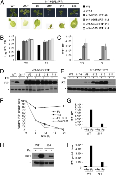
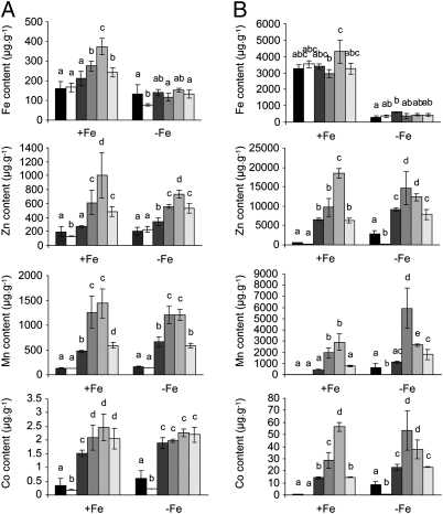
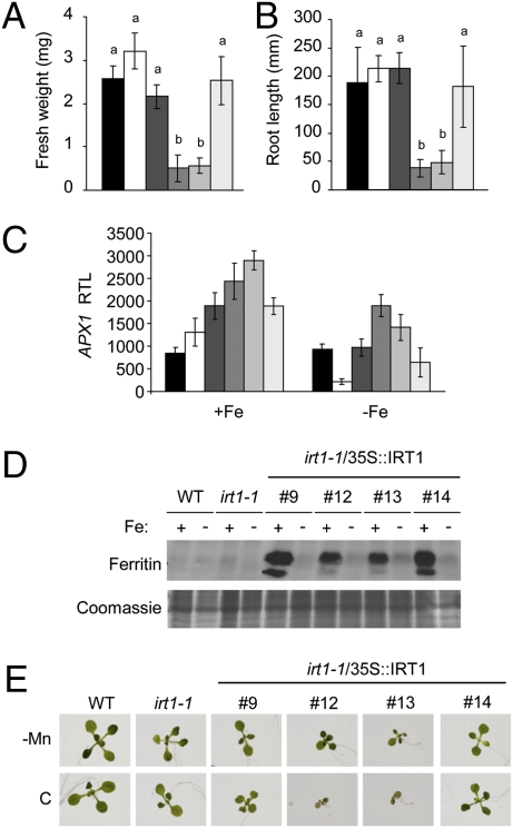
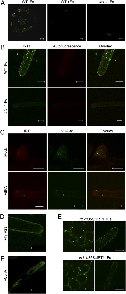
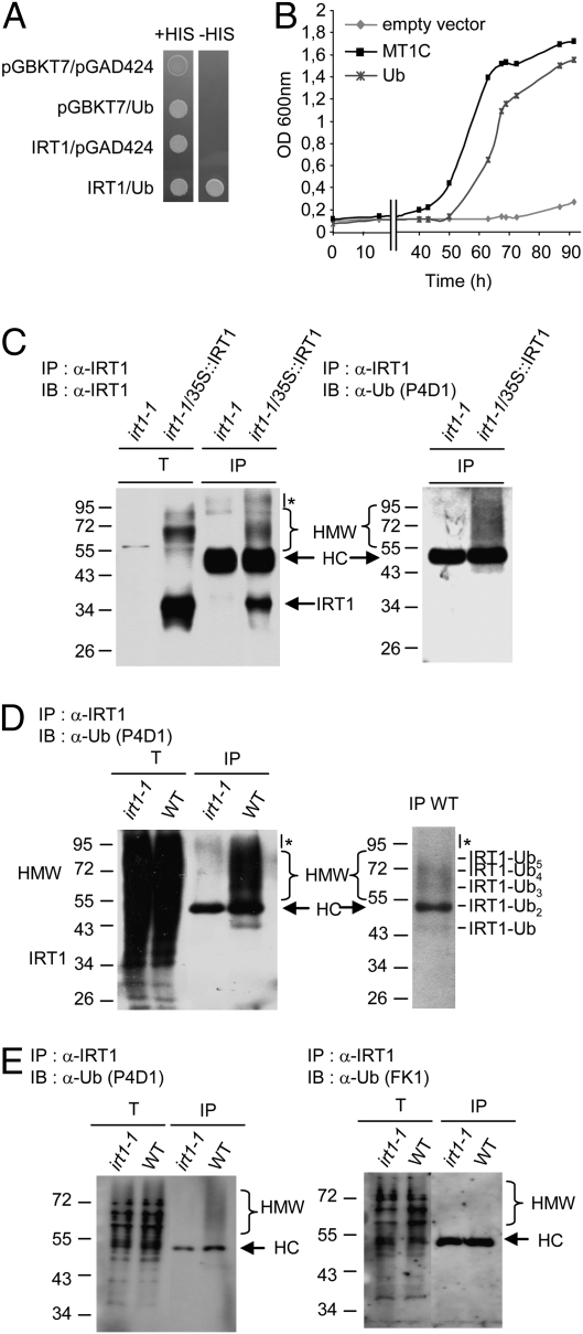
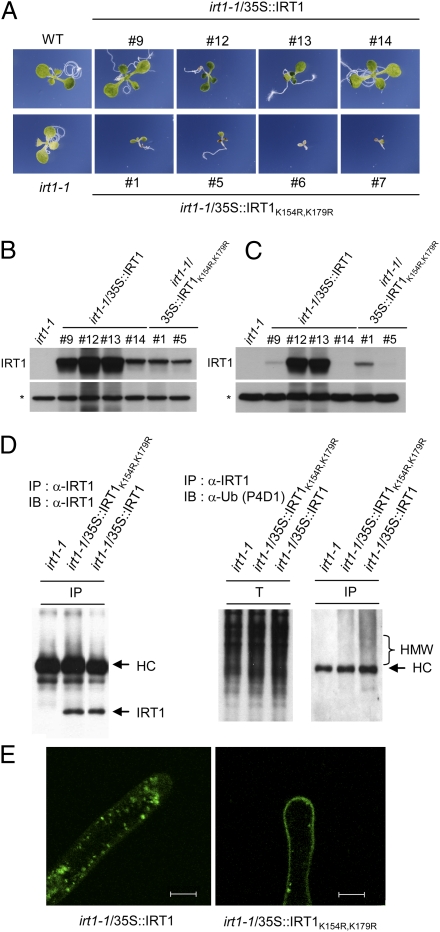
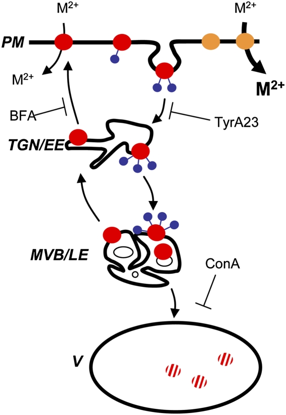
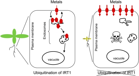
Plants take up iron from the soil using the iron-regulated transporter 1 (IRT1) high-affinity iron transporter at the root surface. Sophisticated regulatory mechanisms allow plants to tightly control the levels of IRT1, ensuring optimal absorption of essential but toxic iron. Here, we demonstrate that overexpression of Arabidopsis thaliana IRT1 leads to constitutive IRT1 protein accumulation, metal overload, and oxidative stress. IRT1 is unexpectedly found in trans-Golgi network/early endosomes of root hair cells, and its levels and localization are unaffected by iron nutrition. Using pharmacological approaches, we show that IRT1 cycles to the plasma membrane to perform iron and metal uptake at the cell surface and is sent to the vacuole for proper turnover. We also prove that IRT1 is monoubiquitinated on several cytosol-exposed residues in vivo and that mutation of two putative monoubiquitination target residues in IRT1 triggers stabilization at the plasma membrane and leads to extreme lethality. Together, these data suggest a model in which monoubiquitin-dependent internalization/sorting and turnover keep the plasma membrane pool of IRT1 low to ensure proper iron uptake and to prevent metal toxicity. More generally, our work demonstrates the existence of monoubiquitin-dependent trafficking to lytic vacuoles in plants and points to proteasome-independent turnover of plasma membrane proteins.


Conflict of interest statement

The authors declare no conflict of interest.

Figures

Fig. 1.
Fig. 1.
Overexpression of IRT1 leads to constitutive accumulation of IRT1 protein, regardless of iron supply. (A) Phenotype of plants constitutively expressing IRT1. Real-time quantitative RT-PCR monitoring of IRT1 transcript accumulation in roots (B) and leaves (C) of WT, irt1-1, and IRT1-overexpressing plants. Experiments were performed on RNA extracted from 10-d-old plants transferred for 3 d on iron-sufficient (+Fe) or iron-deficient (−Fe) conditions. Four independent irt1-1/35S::IRT1 transgenic lines are shown. The results for IRT1 expression in roots are shown as a logarithm of relative transcript levels to visualize IRT1 induction by iron starvation in WT roots. RTL, relative transcript level. Error bars indicate SD. Western blot analyses monitoring of IRT1 protein levels in roots (D) and leaves (E) of WT, irt1-1, and IRT1 overexpressors. The nonspecific band indicated with an asterisk serves as a loading control. (F) Half-life of endogenous IRT1 protein. Western blot analyses were performed using an anti-IRT1 antibody on total root protein extracts from WT −Fe plants transferred to +Fe or −Fe conditions in the presence or absence of CHX. (G) Quantitative RT-PCR monitoring of IRT1 expression in WT and fit-1 roots. Experiments were performed using RNA extracted from roots of 10-d-old plants transferred for 3 d in +Fe or −Fe conditions. The result from a representative experiment is shown. (H) IRT1 protein accumulation profile in roots from WT and fit-1 plants grown in the same conditions as in G. The nonspecific band indicated with an asterisk serves as a loading control. (I) Quantification of the chemiluminescence signals from experiments presented in H.
Fig. 2.
Fig. 2.
IRT1 overexpression leads to metal accumulation independent of iron nutrition. Metal content was determined by inductively coupled plasma mass spectrometry (ICP-MS) on leaves (A) and roots (B) from WT (black), irt1-1 (white), irt1-1/35S::IRT1#9 (dark gray), irt1-1/35S::IRT1#12 (medium dark gray), irt1-1/35S::IRT1#13 (medium light gray), and irt1-1/35S::IRT1#14 (light gray) transgenic lines transferred to iron-sufficient (+Fe) or iron-deficient (−Fe) conditions for 5 d. Results are presented as mean ± SD (n = 3). Statistical differences were calculated by one-way ANOVA. Different letters indicate means that were statistically different by Tukey's multiple testing method (P < 0.05) for genotypes within a given growth condition (+Fe or −Fe).
Fig. 3.
Fig. 3.
IRT1-dependent metal accumulation affects growth and triggers oxidative stress responses. (A) Biomass measurements. Fresh weight was measured on 10-d-old WT (black), irt1-1 (white), irt1-1/35S::IRT1#9 (dark gray), irt1-1/35S::IRT1#12 (medium dark gray), irt1-1/35S::IRT1#13 (medium light gray), and irt1-1/35S::IRT1#14 (light gray) transgenic plants grown in standard conditions. Results are presented as mean ± SD (n = 10). Statistical differences were calculated by one-way ANOVA. Different letters indicate means that were statistically different by Tukey's multiple testing method (P < 0.05). (B) Root length measurements in WT, irt1-1, and plants overexpressing IRT1. Results are presented as mean ± SD (n = 15). Statistical differences were calculated by one-way ANOVA. Different letters indicate means that were statistically different by Tukey's multiple testing method (P < 0.05). (C) Quantitative RT-PCR analyses monitoring APX1 expression in leaves of WT, irt1-1, and irt1-1/35S::IRT1 transgenic lines. Experiments were performed using 10-d-old plants transferred for 3 d in iron-sufficient (+Fe) or iron-deficient (−Fe) conditions. Error bars represent SD. (D) Ferritin accumulation profile in leaves from WT, irt1-1, and IRT1 overexpressors determined by Western blot analysis. Plants were grown as in C. (E) Phenotype of IRT1 overexpressors in manganese-deficient conditions. WT, irt1-1, and irt1-1/35S::IRT1 transgenic plants were grown in standard conditions (C) or in manganese-poor (−Mn) medium for 12 d. Representative plants from each genotype are shown.
Fig. 4.
Fig. 4.
IRT1 protein dynamically localizes to the early endosomes. (A) Immunofluorescence using anti-IRT1 antibody on root cross-section of WT and irt1-1 plants. (B) Whole-mount immunolocalization monitoring IRT1 protein in root hairs of WT and irt1-1 null mutant. The cell wall autofluorescence is shown in the red channel. (C) Colocalization of IRT1 with VHA-a1. Iron-starved VHA-a1–GFP transgenic plants were subjected to whole-mount immunolocalization using anti-GFP (Alexa488, green) and anti-IRT1 (Cy3, red) antibodies in the presence of mock (Upper) or BFA (Lower) treatment. Colocalization of IRT1 and VHA-a1 is shown in yellow in the overlay. (D) Immunolocalization of IRT1 in iron-starved WT plants following TyrA23 treatment. (E) Immunolocalization of IRT1 in root cross-sections (Left) and root hairs (Right) of irt1-1/35S::IRT1 plants grown in iron-sufficient (+Fe, Upper) or iron-deficient (−Fe, Lower) conditions. (F) Immunolocalization of IRT1 iron-starved WT plants after ConA treatment. (Scale bar = 10 μm.)
Fig. 5.
Fig. 5.
Monoubiquitin-dependent trafficking and turnover of IRT1. (A) Yeast two-hybrid test monitoring interaction between the IRT1 loop and ubiquitin. The corresponding empty vectors were used as controls. The interaction is revealed by the activation of HIS3 transcription and growth on −HIS medium. (B) Functional cadmium sensitivity test in yeast. Yeast cells expressing IRT1 were transformed with empty vector, polyubiquitination, or the metallothionein MT1C-positive control and were grown in the presence of 0.2 μM cadmium. Yeast growth was followed by measuring the optical density over 96 h. (C) In vivo ubiquitination analyses of IRT1. Immunoprecipitation was performed using an anti-IRT1 antibody on solubilized root protein extracts (T) from iron-replete irt1-1/35S::IRT1 or irt1-1 plants and subjected to immunoblotting with an anti-IRT1 antibody (Left) or the antiubiquitin (P4D1) antibody that recognizes monoubiquitination and polyubiquitination (Right). IB, immunoblotting; IP, immunoprecipitation. The heavy chain of IRT1 antibody and the high-molecular-weight smear specific for IRT1 immunoprecipitates are shown. HC, heavy chain; HMW, high molecular weight. The asterisk indicates aspecific signals observed in both irt1-1 and irt1-1/35S::IRT1 lanes. (D) Ubiquitination analyses of endogenous IRT1. IRT1-immunoprecipitated fractions from WT and irt1-1 iron-deficient plants were subjected to Western blot analysis with the antiubiquitin (P4D1) antibody. (Right) Lower exposure, with several bands corresponding to ubiquitinated forms of IRT1, each migrating ∼9 kDa apart. (E) IRT1 monoubiquitination analyses. IRT1 immunoprecipitates were subjected to Western blot analysis with both the antimonoubiquitination/polyubiquitination-specific (P4D1, Left) and polyubiquitination-specific (FK1, Right) antibodies.
Fig. 6.
Fig. 6.
Monoubiquitination of two lysine residues controls IRT1 localization and degradation. (A) Phenotypic analysis of seedlings from independent transgenic lines expressing IRT1 and IRT1K154R,K179R under the control of 35S promoter. Western blot analyses monitoring IRT1 protein accumulation in roots (B) and shoots (C) of irt1-1/35S::IRT1 and irt1-1/35S::IRT1K154R,K179R transgenic lines. (D) In vivo ubiquitination profile of IRT1. IRT1 immunoprecipitates from irt1-1, irt1-1/35S::IRT1, and irt1-1/35S::IRT1K154R,K179R plants were subjected to Western blot analysis with the anti-IRT1 (Left) and the antiubiquitin (P4D1, Right) antibodies. IB, immunoblotting; IP, immunoprecipitation. (E) Whole-mount immunolocalization analyses of IRT1 in irt1-1/35S::IRT1 and irt1-1/35S::IRT1K154R,K179R plants. (Scale bar = 10 μm.)
Fig. 7.
Fig. 7.
Diagram illustrating the dynamics of IRT1 protein in the cell. The continuous monoubiquitination-dependent cycling of IRT1 (red circles) between the plasma membrane (PM) and early endosomes (TGN/EE) controls its subcellular distribution and transport of divalent metals (M2+). A fraction of monoubiquitinated endocytosed IRT1 protein is not recycled to the plasma membrane and is constantly sent for degradation in the vacuole (V) via multivesicular bodies (MVBs/LEs), thereby controlling IRT1 protein total levels. Inhibition of endocytosis (TyrA23), recycling to the plasma membrane (BFA), degradation in the vacuole (ConA), and mutation of residues K154 and K179 (orange circles) interfere with intracellular distribution of IRT1.
Fig. P1.
Fig. P1.
Biological relevance of ubiquitin-dependent control of IRT1 localization. Plants take up iron and metals from the soil using the IRT1 root iron transporter (red). (Left) Our study demonstrates that IRT1 is localized to endosomes but transiently goes at the plasma membrane of root hair cells to take up metals from the soil. The endosomal localization of IRT1 requires its monoubiquitination. (Right) When a mutant version of IRT1 that does not undergo ubiquitination is expressed in plants, IRT1 is readily found at the cell surface and takes up an excess of metals. Such plants develop major oxidative stress, leading to the appearance of leaf necrotic spots, and show severe growth reduction or even lethality.

References

    1. Curie C, Briat JF. Iron transport and signaling in plants. Annu Rev Plant Biol. 2003;54:183–206. - PubMed
    1. Briat JF, et al. Cellular and molecular aspects of iron metabolism in plants. Biol Cell. 1995;84:69–81.
    1. Vert G, et al. IRT1, an Arabidopsis transporter essential for iron uptake from the soil and for plant growth. Plant Cell. 2002;14:1223–1233. - PMC - PubMed
    1. Henriques R, et al. Knock-out of Arabidopsis metal transporter gene IRT1 results in iron deficiency accompanied by cell differentiation defects. Plant Mol Biol. 2002;50:587–597. - PubMed
    1. Colangelo EP, Guerinot ML. The essential basic helix-loop-helix protein FIT1 is required for the iron deficiency response. Plant Cell. 2004;16:3400–3412. - PMC - PubMed

Publication types