. 2011 Jun 14;108(24):9875-80.
doi: 10.1073/pnas.1100816108. Epub 2011 May 31.

Multilineage-differentiating stress-enduring (Muse) cells are a primary source of induced pluripotent stem cells in human fibroblasts

Affiliations

Multilineage-differentiating stress-enduring (Muse) cells are a primary source of induced pluripotent stem cells in human fibroblasts

Shohei Wakao et al. Proc Natl Acad Sci U S A. .

Abstract

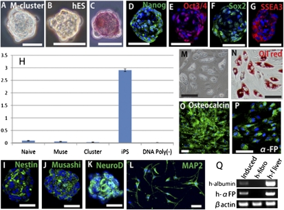
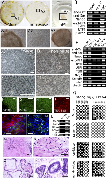
The stochastic and elite models have been proposed for the mechanism of induced pluripotent stem (iPS) cell generation. In this study we report a system that supports the elite model. We previously identified multilineage-differentiating stress-enduring (Muse) cells in human dermal fibroblasts that are characterized by stress tolerance, expression of pluripotency markers, self-renewal, and the ability to differentiate into endodermal-, mesodermal-, and ectodermal-lineage cells from a single cell. They can be isolated as stage-specific embryonic antigen-3/CD105 double-positive cells. When human fibroblasts were separated into Muse and non-Muse cells and transduced with Oct3/4, Sox2, Klf4, and c-Myc, iPS cells were generated exclusively from Muse cells but not from non-Muse cells. Although some colonies were formed from non-Muse cells, they were unlike iPS cells. Furthermore, epigenetic alterations were not seen, and some of the major pluripotency markers were not expressed for the entire period during iPS cell generation. These findings were confirmed further using cells transduced with a single polycistronic virus vector encoding all four factors. The results demonstrate that in adult human fibroblasts a subset of preexisting adult stem cells whose properties are similar in some respects to those of iPS cells selectively become iPS cells, but the remaining cells make no contribution to the generation of iPS cells. Therefore this system seems to fit the elite model rather than the stochastic model.


Conflict of interest statement

The authors declare no conflict of interest.

Figures

Fig. 1.
Fig. 1.
Muse cells in human dermal fibroblasts. (A and B) Examples of flow cytometry analysis of SSEA-3 (A) and CD105 (B) expression in naive human cultured fibroblasts. (CF) Flow cytometry analysis of SSEA-3+ cells. Because naive fibroblasts showed no reactivity to NG2 (C), CD34 (D), von Willebrand factor (vWF) (E), or CD31 (F), SSEA-3+ cells were negative for these markers also. (GI) A small percentage of naive fibroblasts were positive for CD117, CD146, and CD271. However, these cells were SSEA-3. (J) RT-PCR shows that, although SSEA-3 cells (non-Muse cells) showed weak signals for Snai1 and Slug, SSEA-3+ cells (Muse cells) were negative for all markers. Human embryo (embryo) served as the positive control. (K) (Left) H&E staining of adult human skin and (Right) immunohistochemistry of SSEA-3+ cells in boxed areas 1–3. Positive cells were observed in the connective tissue distributed in dermis (1) and sweat gland (2) (Upper Right) and around small blood vessels (3) (Lower Right). (L and M) SSEA-3 immunohistochemistry of adult human skin combined with counterstaining (hematoxylin staining) in the neighboring section. (Scale bars, 50 μm.)
Fig. 2.
Fig. 2.
Character of Muse cells and M-clusters. (A) M-cluster formed in single-cell suspension culture. (B) Cell cluster formed by hESC (hES) in suspension culture. (C) ALP staining of an M-cluster. (DG) Immunocytochemistry of (D) Nanog, (E) Oct3/4, (F) Sox2, and (G) SSEA-3 in M-clusters. (H) Telomerase activity in naive fibroblasts (Naive), Muse cells (Muse), M-clusters (Cluster), and Fibro-iPSs (iPS). DNA Poly(−) represents a negative control. (IQ) Controlled differentiation of Muse cells. Neural induction produced spheres that expressed nestin (I), Musashi (J), and NeuroD (K). (L) MAP-2+ cells after differentiation. (M and N) Induced adipocytes contained lipid droplets (M) that could be stained with oil red O (oil red) (N). Induced osteoblasts expressed osteocalcin (O). (P and Q) Induced hepatocytes were positive for human α-FP (P). (Q) RT-PCR analysis after hepatocyte induction (Induced) detected human albumin (h-albumin) and human α-FP (h-αFP). Human naive fibroblasts (h-fibro) served as a negative control, and human fetal liver (h-f liver) served as a positive control. (Scale bars: AG, and L, 30 μm; IK and M–O, 50 μm.)
Fig. 3.
Fig. 3.
Generation of iPS cells from Muse cells. (A) (Upper)After 30-d culture on MEFs, colonies derived from Muse cells were TRA-1–81+, whereas those derived from non-Muse cells were TRA-1–81. hESCs (hES) were the positive control. (Lower) Enlarged views of boxed areas 1–3 are shown in A1–A3. (B) RT-PCR for endogenous Oct3/4 (end-Oct), endogenous Sox2 (end-Sox), Nanog, endogenous Klf4 (end-Klf4), Rex1, and UTF1 Muse and non-Muse (non-M) cells after 30-d culture on MEFs. hESCs (hES) were the positive control. (C and D) The appearance of Muse-iPS cells (C and C1) and non-Muse colony (D and D1) after colony pickup. (E) RT-PCR for endogenous Oct3/4 (end-Oct), endogenous Sox2 (end-Sox), Nanog, endogenous Klf4 (end-Klf4), Rex1, UTF1, TERT, Abcg2, Dnmt3b, and Cdx2 in Muse-iPS cells (two representative clones: M iPS-1 and M iPS -2) and non-Muse colonies (two representative clones: non–M-1 and non–M-2) after colony pickup. (FI) Immunocytochemistry of (F) Nanog, (G) Oct3/4, (H) Sox2, and (I) TRA-1–81 in Muse-iPS cells and immunocytochemistry of cells expanded from Muse-iPS cell–derived embryoid bodies showing the expression of (J) SMA (α-SMA; red) and α-FP; (green) and (K) neurofilament (NF; green). (L) RT-PCR of differentiation markers in untreated Muse-iPS cells (Undiff) and embryoid body derived from Muse-iPS cells (Diff). (MP) H&E staining of teratoma (M) that formed in the testes of immunodeficient mice 12 wk after injection of Muse-iPS cells. Mesodermal (N), endodermal (O), and ectodermal (P) tissues were recognized within the teratoma. (Q) Bisulfite sequencing of the promoter regions of Nanog and Oct3/4 in Muse cells, Muse-iPS cells, non-Muse cells, and non-Muse colonies (non-M colony). Empty and filled circles represent unmethylated and methylated cytosines, respectively. (Scale bar: C and D, 100 μm; C1 and D1, 50 μm; FK, 50 μm; MP, 100 μm.)

References

    1. Kørbling M, Estrov Z. Adult stem cells for tissue repair - a new therapeutic concept? N Engl J Med. 2003;349:570–582. - PubMed
    1. Thomas ED. Landmarks in the development of hematopoietic cell transplantation. World J Surg. 2000;24:815–818. - PubMed
    1. Thomson JA, et al. Embryonic stem cell lines derived from human blastocysts. Science. 1998;282:1145–1147. - PubMed
    1. Takahashi K, et al. Induction of pluripotent stem cells from adult human fibroblasts by defined factors. Cell. 2007;131:861–872. - PubMed
    1. Sullivan GJ, Bai Y, Fletcher J, Wilmut I. Induced pluripotent stem cells: Epigenetic memories and practical implications. Mol Hum Reprod. 2010;16:880–885. - PubMed

Publication types

MeSH terms

Substances

LinkOut - more resources