. 2011 Jun 14;108(24):9945-50.
doi: 10.1073/pnas.1100890108. Epub 2011 May 31.

In vivo diagnosis of murine pancreatic intraepithelial neoplasia and early-stage pancreatic cancer by molecular imaging

Affiliations

In vivo diagnosis of murine pancreatic intraepithelial neoplasia and early-stage pancreatic cancer by molecular imaging

Stefan Eser et al. Proc Natl Acad Sci U S A. .

Abstract

Pancreatic ductal adenocarcinoma (PDAC) is a fatal disease with poor patient outcome often resulting from late diagnosis in advanced stages. To date methods to diagnose early-stage PDAC are limited and in vivo detection of pancreatic intraepithelial neoplasia (PanIN), a preinvasive precursor of PDAC, is impossible. Using a cathepsin-activatable near-infrared probe in combination with flexible confocal fluorescence lasermicroscopy (CFL) in a genetically defined mouse model of PDAC we were able to detect and grade murine PanIN lesions in real time in vivo. Our diagnostic approach is highly sensitive and specific and proved superior to clinically established fluorescein-enhanced imaging. Translation of this endoscopic technique into the clinic should tremendously improve detection of pancreatic neoplasia, thus reforming management of patients at risk for PDAC.


Conflict of interest statement

The authors declare no conflict of interest.

Figures

Fig. 1.
Fig. 1.
Cathepsins are highly expressed in preinvasive mPanIN lesions and early-stage PDAC. (A) mRNA expression profiling of pancreatic tissues from wild-type controls (WT), mice with caerulein-induced pancreatitis, and Ptf1aCre/+;LSL-KrasG12D/+ mice bearing mPanINs and PDAC, respectively. RNA was isolated from tissue specimens, labeled, and hybridized onto a GeneChip Mouse Genome 430 2.0 array (Affymetrix). Expression levels of cathepsin proteases are shown as log-fold change in row-Z scale (ctsb, cathepsin B; ctsh, cathepsin H; ctsl, cathepsin L; ctss, cathepsin S). (B) Paraffin-embedded pancreatic tissue sections from wild-type mice, caerulein-treated mice with pancreatitis, and Ptf1aCre/+;LSL-KrasG12D/+ mice with mPanIN lesions and PDAC were stained for ctsb, ctsh, ctsl, and ctss.
Fig. 2.
Fig. 2.
Specific activation of a cathepsin-sensitive NIRF probe in mPanIN lesions and PDAC. (A) Near-infrared (NIRF) imaging of pancreatic tissues from Ptf1aCre/+;LSL-KrasG12D/+ mice bearing mPanIN lesions and PDAC. Wild-type (WT) littermates and mice with caerulein-induced acute pancreatitis served as controls. Tissues were harvested 24 h after i.v. injection of the cathepsin-activatable NIRF probe. Cryosections were then scanned with a planar Odyssey near-infrared reader at 680 nm for visualization of the NIRF probe. The NIRF-probe signal is shown in red. Bright field imaging (green color) to show morphology was performed at 800 nm. (B) Histological analysis of the serial section of the scanned PDAC specimen depicted in A shows necrosis next to viable tumor, correlating with the NIRF-probe signal (magnification: left, 2×; right, 200×). (C) Quantification of fluorescence intensity in scanned cryosections shown in A using Odyssey software. (D) Confocal lasermicroscopic imaging of activated cathepsin-sensitive NIRF probes in cryosections from normal pancreas, caerulein-induced pancreatitis, low- and high-grade mPanIN lesions, and PDAC (magnification, 200×). The signal from the activated NIRF probe is depicted in red. E-cadherin counterstain displays morphology (green).
Fig. 3.
Fig. 3.
Early detection of preinvasive mPanIN lesions and PDAC in vivo by flexible confocal lasermicroscopy. (A, Upper) In vivo imaging of all grades of mPanIN lesions and PDAC in Ptf1aCre/+;LSL-KrasG12D/+ mice and normal pancreas in littermate controls on a cellular level using CFL and a cathepsin-activatable NIRF probe. The cathepsin-sensitive NIRF-probe signal increases with progression of mPanINs to PDAC and is almost absent in normal pancreatic tissue. Additionally, the NIRF-probe signal provides valuable information about the morphology of the lesion on a cellular level. White asterisks in the PDAC image indicate two ductal structures characteristic of PDAC ín CFL. (Lower) The corresponding matched H&E stained histological sections are shown. Black asterisks mark the ductal structures detected by CFL in the PDAC specimen. (B) Ex vivo whole organ scans of the pancreas with stomach and duodenum of control and 3-, 6-, and 12-mo-old Ptf1aCre/+;LSL-KrasG12D/+ animals bearing mPanIN lesions (3- and 6-mo-old mice) or PDAC (12-mo-old mouse) on a planar Odyssey near-infrared scanner. NIRF-probe signal imaged at 680 nm is shown in red. Bright field images scanned at 800 nm appear in green.
Fig. 4.
Fig. 4.
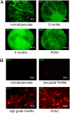
Real-time dual wavelength in vivo imaging of cathepsin-activatable NIRF probes and the nonspecific vascular contrast agent fluorescein to detect mPanIN lesions and PDAC. (A) Flexible confocal lasermicroscopic in vivo imaging of Ptf1aCre/+;LSL-KrasG12D/+ and wild-type control mice after laparatomy using the nonspecific vascular contrast agent fluorescein (green color). (Scale bar, 50 μm.) Morphology and extent of fibrosis is shown in normal pancreas, pancreas with low- (3 mo), and with high-grade mPanIN lesions (6 mo) and PDAC. (B) CFL imaging of cathepsin activity of Ptf1aCre/+;LSL-KrasG12D/+ mice and wild-type littermate controls. (Scale bar, 20 μm.) Pictures shown depict normal pancreas, low- and high-grade mPanIN lesions, and PDAC.

References

    1. Schneider G, Siveke JT, Eckel F, Schmid RM. Pancreatic cancer: Basic and clinical aspects. Gastroenterology. 2005;128:1606–1625. - PubMed
    1. Furukawa H, et al. Clinicopathologic features of small pancreatic adenocarcinoma. A collective study. Cancer. 1996;78:986–990. - PubMed
    1. Agarwal B, Correa AM, Ho L. Survival in pancreatic carcinoma based on tumor size. Pancreas. 2008;36:e15–e20. - PubMed
    1. Shimada K, Sakamoto Y, Sano T, Kosuge T, Hiraoka N. Reappraisal of the clinical significance of tumor size in patients with pancreatic ductal carcinoma. Pancreas. 2006;33:233–239. - PubMed
    1. Shimizu Y, Yasui K, Matsueda K, Yanagisawa A, Yamao K. Small carcinoma of the pancreas is curable: New computed tomography finding, pathological study and postoperative results from a single institute. J Gastroenterol Hepatol. 2005;20:1591–1594. - PubMed

Publication types

MeSH terms