. 2011 Nov;19(11):1144-51.
doi: 10.1038/ejhg.2011.97. Epub 2011 Jun 1.

17p13.3 microduplications are associated with split-hand/foot malformation and long-bone deficiency (SHFLD)

Affiliations

17p13.3 microduplications are associated with split-hand/foot malformation and long-bone deficiency (SHFLD)

Christine M Armour et al. Eur J Hum Genet. 2011 Nov.

Abstract

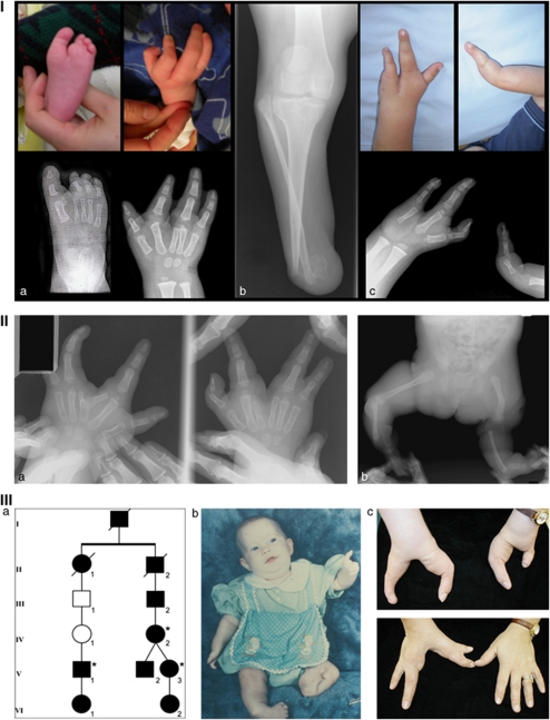
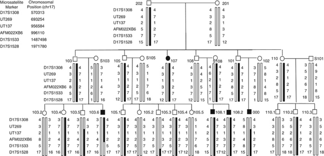
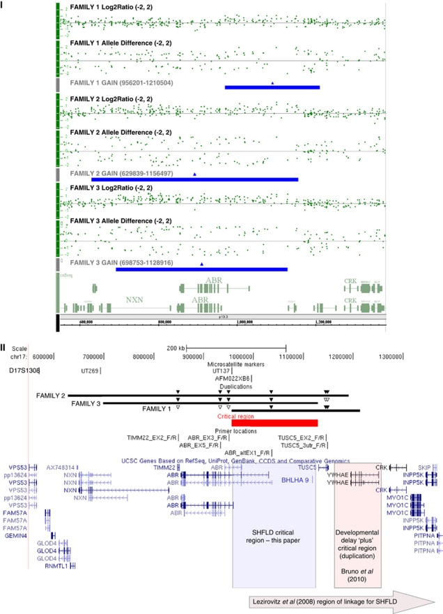
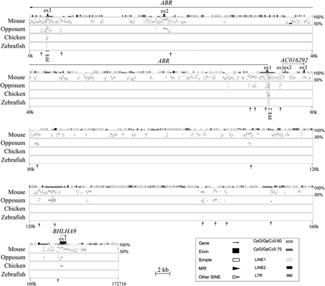
Split-hand/foot malformation with long-bone deficiency (SHFLD) is a relatively rare autosomal-dominant skeletal disorder, characterized by variable expressivity and incomplete penetrance. Although several chromosomal loci for SHFLD have been identified, the molecular basis and pathogenesis of most SHFLD cases are unknown. In this study we describe three unrelated kindreds, in which SHFLD segregated with distinct but overlapping duplications in 17p13.3, a region previously linked to SHFLD. In a large three-generation family, the disorder was found to segregate with a 254 kb microduplication; a second microduplication of 527 kb was identified in an affected female and her unaffected mother, and a 430 kb microduplication versus microtriplication was identified in three affected members of a multi-generational family. These findings, along with previously published data, suggest that one locus responsible for this form of SHFLD is located within a 173 kb overlapping critical region, and that the copy gains are incompletely penetrant.


Figures

Figure 1
Figure 1
Panel I: clinical findings in family 1. (a) Hand and foot findings in the proband. (b) Tibial hypoplasia in individual 107. (c) Monodactyly and oligodactyly with split hand in individual S108.2 who also has tibial hypoplasia (not shown). Pedigree of Family 1 can be found in Figure 2. Panel II: clinical findings in the isolated affected individual from family 2 showing hand malformations (a) including six metacarpals, bifid metacarpal with split hand and (b) tibial aplasia. Radiographs courtesy of Shriners Hospital for Children, Greenville, SC. Panel III: partial pedigree and clinical findings in family 3. (a) Relationships between the individuals from whom samples were available (asterisks). (b) Preoperative photograph of individual V-3 showing hand malformations, distal hypoplasia of right tibia, aplasia of left tibia, and clubfeet. (c) Hand findings in individuals V-3 (upper) and IV-2 (lower) as adults. Individual V-3 was born with only a thumb on the left hand (b), and the lateral digit shown here is a transplanted hallux.
Figure 2
Figure 2
Pedigree with haplotypes of the region on 17p13.3. Circles (○)=females and squares (□)=males. Solid symbols=affected; unshaded symbols=unaffected. Legend to the left of each generation indicates microsatellite marker used and list (top left) indicates genomic co-ordinates (chr 17) of each. The black portion of each haplotype bar indicates haplotype that segregates with the duplication. Individuals 000 and S108 were found to carry the duplication by array (corresponding to region spanning markers UT137 and AFM022XB6). qPCR confirmed array results and also confirmed that individuals 107, 108.2, 103.5, and 103.6 carry the duplication. Three offspring of individuals 101 and 102 and their children did not participate; two sons (reportedly unaffected) with eight children between them (sexes unknown, also reportedly unaffected), and one daughter (reportedly with a ‘short toe') and her five children (reportedly unaffected).
Figure 3
Figure 3
Panel I: Affymetrix Genome-wide SNP 6.0 microarray results (NCBI36/hg18 build). Chromosome 17p13.3 (550000-1380000) showing the copy-number gain in representative affected family members from families 1 (top), 2 (middle), and 3 (bottom). Data for each patient are represented by log2 ratio and allele calls, and the region of copy gain is denoted as a solid bar below the data for each patient. Panel II: relevant area of chromosome 17 from UCSC (http://genome.ucsc.edu/) showing annotated genes., Beneath scale co-ordinates at top are the microsatellites used with their respective positions. Marker D17S1528 is located more centromeric than can be shown in the figure. Duplications as indicated (genomic co-ordinates in panel I). Overlapping area extends over 172 715 nucleotides and is indicated by solid line. Solid and open triangles above each of the families' duplications indicate confirmation of increased and normal dosage, respectively, by qPCR (qPCR data in Supplementary Figure and Table). SHFLD critical region indicated by labelled shaded box. Critical region described by Bruno et al involving YWHAE indicated as labelled. Area of linkage reported by Lezirovitz et al indicated by labelled shaded arrow at bottom.
Figure 4
Figure 4
Cross-species comparative sequence analysis of the SHFLD critical region. The graph compares the percent identity between human genomic sequence of 173 kb with corresponding sequences from mouse, opossum, chicken, and zebrafish. Aligned segments that show over 50% percent of identity are plotted as a series of dots (stippling) according to their length and position within the human sequence. The human sequence is shown on the horizontal axis and the percent identities with non-human sequences are shown on the vertical axis. Annotations correspond to the human sequence (top of plot). Gene sequences are represented by solid-black arrows indicating the direction of transcription, and exons are indicated by black boxes. ABR and BHLHA9 are known protein-coding genes; AC016292.1 is a putative processed transcript. Shaded regions depict putative functional elements, PFE1 and PFE2, which are conserved between human, mouse, opossum, and chicken. Grey vertical arrows depict regions of conservation between human, mouse, and chicken. The analysis also detects and annotates various repetitive sequences in the reference sequence (human). These include simple repeats, long terminal repeats (LTRs), mammalian interspersed elements (MIRs), long interspersed nuclear elements (LINEs), and short interspersed nuclear elements (SINEs). Such sequences are commonly present in multiple copies per genome and are excluded from sequence comparisons. For further details on PIP analyses, see http://bio.cse.psu.edu/pipmaker.

References

    1. Majewski F, Kuster W, ter Haar B, Goecke T. Aplasia of tibia with split-hand/split-foot deformity. Report of six families with 35 cases and considerations about variability and penetrance. Hum Genet. 1985;70:136–147. - PubMed
    1. Basel D, Kilpatrick MW, Tsipouras P. The expanding panorama of split hand foot malformation. Am J Med Genet A. 2006;140:1359–1365. - PubMed
    1. Ugur SA, Tolun A. Homozygous WNT10b mutation and complex inheritance in split-Hand/Foot malformation. Hum Mol Genet. 2008;17:2644–2653. - PubMed
    1. Celli J, Duijf P, Hamel BC, et al. Heterozygous germline mutations in the p53 homolog p63 are the cause of EEC syndrome. Cell. 1999;99:143–153. - PubMed
    1. Kjaer KW, Hansen L, Schwabe GC, et al. Distinct CDH3 mutations cause ectodermal dysplasia, ectrodactyly, macular dystrophy (EEM syndrome) J Med Genet. 2005;42:292–298. - PMC - PubMed

Publication types

Substances

Supplementary concepts