. 2010 Jun;27(2):148-59.
doi: 10.1055/s-0030-1253513.

Percutaneous treatment of intervertebral disc herniation

Affiliations

Percutaneous treatment of intervertebral disc herniation

Xavier Buy et al. Semin Intervent Radiol. 2010 Jun.

Abstract

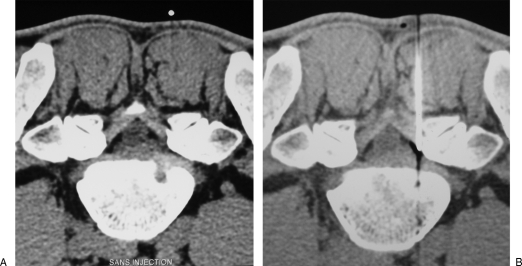
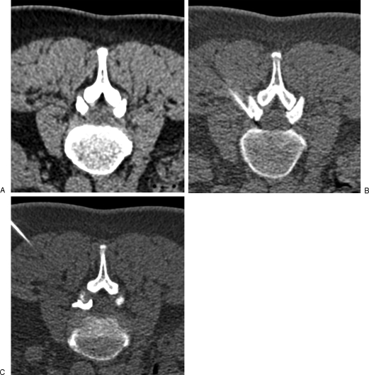
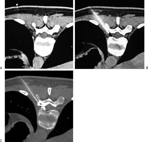
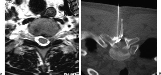
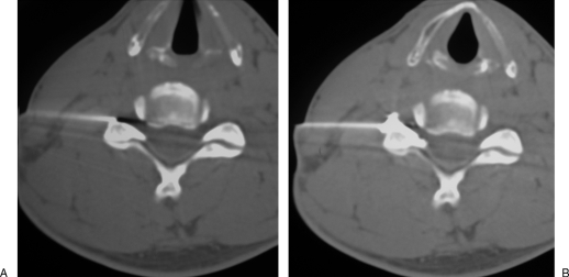
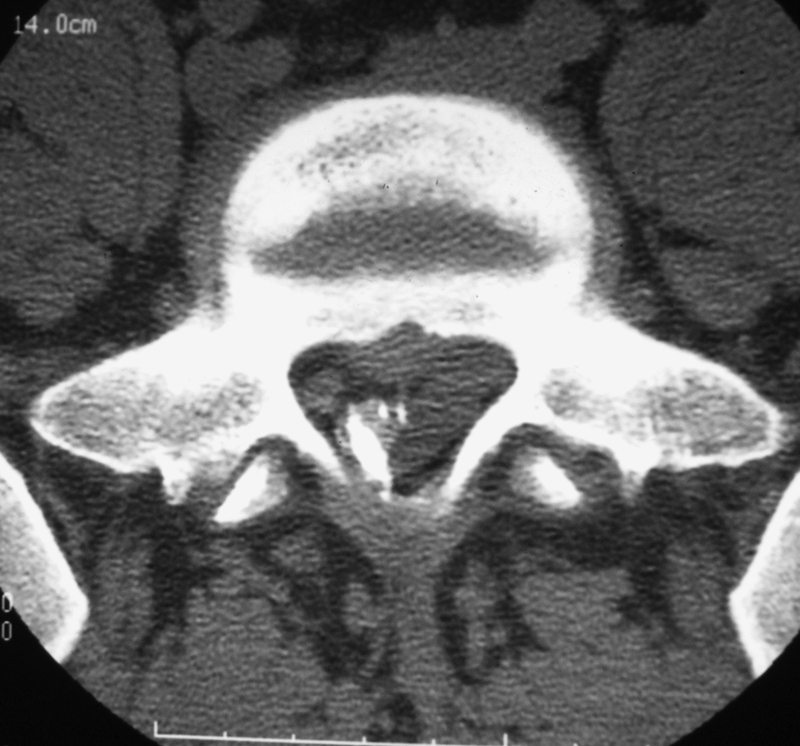
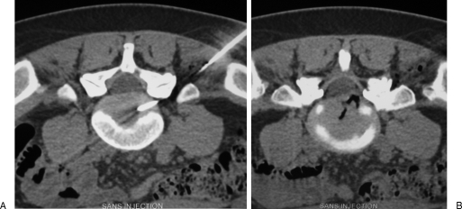
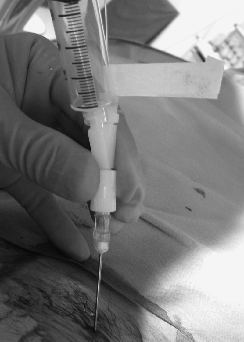
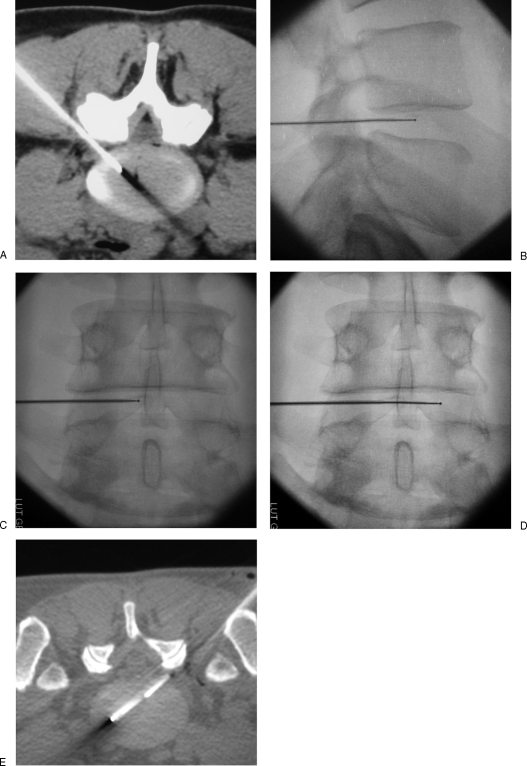
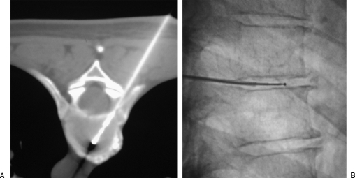
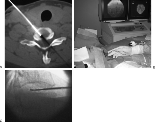
Interventional radiology plays a major role in the management of symptomatic intervertebral disc herniations. In the absence of significant pain relief with conservative treatment including oral pain killers and anti-inflammatory drugs, selective image-guided periradicular infiltrations are generally indicated. The precise control of needle positioning allows optimal distribution of steroids along the painful nerve root. After 6 weeks of failure of conservative treatment including periradicular infiltration, treatment aiming to decompress or remove the herniation is considered. Conventional open surgery offers suboptimal results and is associated with significant morbidity. To achieve minimally invasive discal decompression, different percutaneous techniques have been developed. Their principle is to remove a small volume of nucleus, which results in an important reduction of intradiscal pressure and subsequently reduction of pressure inside the disc herniation. However, only contained disc herniations determined by computed tomography or magnetic resonance are indicated for these techniques. Thermal techniques such as radiofrequency or laser nucleotomy seem to be more effective than purely mechanical nucleotomy; indeed, they achieve discal decompression but also thermal destruction of intradiscal nociceptors, which may play a major role in the physiopathology of discal pain. The techniques of image-guided spinal periradicular infiltration and percutaneous nucleotomy with laser and radiofrequency are presented with emphasis on their best indications.

Keywords: Disc herniation; laser; percutaneous nucleotomy; periradicular infiltration; radiofrequency.


Figures

Figure 1
Figure 1
Lumbar epidural infiltration. (A) Axial computed tomography scan shows a right posterolateral disc herniation at L5–S1 level, responsible for sciatic pain. (B) A 22-gauge needle is inserted into the right lateral epidural space; before steroid injection, proper position of the needle tip is confirmed by diffusion of a small air bubble into the epidural space.
Figure 2
Figure 2
Lumbar foraminal infiltration. (A) Axial computed tomography scan shows a left foraminal disc herniation at L2–L3 level, responsible for crural pain. (B) A 22-gauge needle is inserted into the low part of the foramen, behind the nerve root. (C) Contrast injection is performed to confirm appropriate needle position before steroid injection.
Figure 3
Figure 3
Thoracic foraminal infiltration. (A) Axial computed tomography scan shows a left posterolateral disc herniation responsible for intercostal radicular pain. (B) A 22-gauge needle is inserted into the low part of the foramen, keeping behind the foraminal nerve root. (C) Contrast injection confirms the proper diffusion around the nerve root toward the spinal canal, before steroid injection.
Figure 4
Figure 4
Thoracic epidural infiltration. (A) Axial T2-weighted magnetic resonance image shows a left posterolateral disc herniation at T2–T3 level, responsible for axillary radicular pain. (B) Periradicular steroid injection is performed in the lateral epidural space via a posterior approach.
Figure 5
Figure 5
Cervical infiltration. (A) A 22-gauge needle is inserted into the foramen, behind the nerve root, with its tip in close contact with the anterior border of the facet joint, away from the vertebral artery. (B) Before steroid injection, contrast injection is performed to confirm proper distribution around the nerve root, toward the foramen.
Figure 6
Figure 6
Epidural hematoma following epidural periradicular infiltration. Myelo-computed tomography shows a small left posterolateral collection displacing the dural sac (opacified with contrast). The patient remained asymptomatic.
Figure 7
Figure 7
Percutaneous laser lumbar nucleotomy at L5–S1 level. (A) Laser fiber is inserted into the disc coaxially through a bended 18-gauge needle. Close contact with the facet joint is mandatory to avoid the foraminal nerve root. (B) Computed tomography scan shows the vaporization with gas diffusion into the herniation.
Figure 8
Figure 8
Percutaneous laser nucleotomy. Aspiration is applied via the side arm fitting to avoid excessive intradiscal gas trapping and lumbar pain during vaporization.
Figure 9
Figure 9
Percutaneous lumbar radiofrequency nucleoplasty. (A) A 17-gauge introducer needle is positioned via a posterolateral approach at the posterior annulus/nucleus junction. (B, C) Fluoroscopic views show the radiofrequency electrode inserted coaxially into the nucleus, in proximal position. (D, E) Fluoroscopy and computed tomography show the electrode advanced in distal position, in contact with the anterior annulus. Six to 10 Coblation® channels are dug to achieve discal decompression. The tip of the electrode should not touch the vertebral end plates to avoid thermal damage to their cartilage.
Figure 10
Figure 10
Percutaneous thoracic radiofrequency nucleotomy. (A) The electrode in inserted into the disc, parallel to the adjacent vertebral end plates, between the head of the rib and the pedicle. (B) Lateral fluoroscopic view shows the tip of the electrode, away from the vertebral end plates. Due to reduced height of thoracic discs, only lateral Coblation® channels are dug to avoid damage to the vertebral end plates.
Figure 11
Figure 11
Percutaneous cervical radiofrequency nucleotomy. (A, B) A dedicated 19-gauge radiofrequency electrode is inserted into the cervical disc via an anterolateral approach, just medial to the carotid artery. (C) Three spherical voids are created in the disc by rotation of the looped-tip radiofrequency electrode in the nucleus. The electrode should not be advanced beyond the midthird/posterior-third junction of the disc to avoid damage to the neurological structures.

References

    1. Castro I, Santos D P, Christoph DdeH, Landeiro J A. The history of spinal surgery for disc disease: an illustrated timeline. Arq Neuropsiquiatr. 2005;63(3A):701–706. - PubMed
    1. Burke J G, Watson R W, McCormack D, Dowling F E, Walsh M G, Fitzpatrick J M. Intervertebral discs which cause low back pain secrete high levels of proinflammatory mediators. J Bone Joint Surg Br. 2002;84:196–201. - PubMed
    1. Peng B, Hou S, Wu W, Zhang C, Yang Y. The pathogenesis and clinical significance of a high-intensity zone (HIZ) of lumbar intervertebral disc on MR imaging in the patient with discogenic low back pain. Eur Spine J. 2006;15:583–587. - PMC - PubMed
    1. Coppes M H, Marani E, Thomeer R T, Groen G J. Innervation of “painful” lumbar discs. Spine (Phila Pa 1976) 1997;22:2342–2349. discussion 2349–2350. - PubMed
    1. Smith L, Garvin P J, Gesler R M, Jennings R B. Enzyme dissolution of the nucleus pulposus. Nature. 1963;198:1311–1312. - PubMed