

Neuroendocrine neoplasms of the gastrointestinal tract
- PMID: 21629514
- PMCID: PMC3103981
- DOI: 10.3238/arztebl.2011.0305
Neuroendocrine neoplasms of the gastrointestinal tract
Abstract
Background: Gastroenteropancreatic neuroendocrine neoplasms (GEP-NENs) are complex tumors whose incidence is rising and whose treatment requires precise classification and risk stratification.
Method: Selective review of the relevant literature, including recently published guidelines.
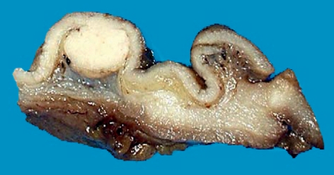
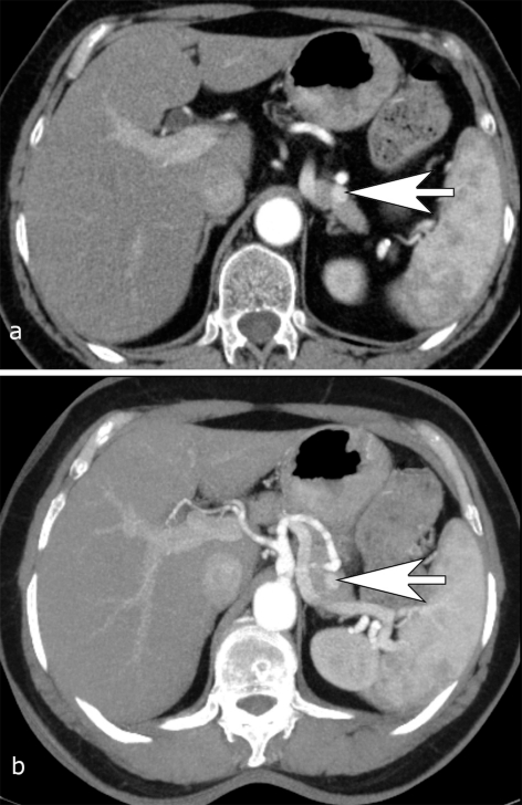
Results: GEP-NENs are initially classified by their degree of histological differentiation and their graded cell proliferation (Ki-67 index). In addition, there are GEP-NEN specific TNM staging protocols. The laboratory assessment includes the measurement of general tumor markers (synaptophysin, chromogranin A) as well as specific ones (hormones). The most important imaging technique for diagnosis is octreotide scintigraphy. The surgical treatment of GEP-NEN is based on oncological resection criteria whose aim is to achieve locally radical resection while preserving as much organ function as possible. Metastases, too, may be amenable to resection. The treatment options for unresectable metastases include radiofrequency ablation and chemoembolization, both of which are palliative methods of reducing tumor volume and hormone production. Other chemotherapeutic and nuclear-medical treatments can be applied depending on the extent of metastatic spread, the proliferation index, and the degree of hormone production by the tumor.
Conclusion: The accurate diagnosis and appropriate treatment of GEP-NET currently gives most patients with this tumor a good prognosis, as long as it is discovered early. Early GEP-NETs have a favorable prognosis. Further advances in the diagnosis and treatment of this disease may result from structural changes in patient care, including the establishment of NET centers.
Figures


Comment in
-
Correspondence (letter to the editor): Incomplete picture.Dtsch Arztebl Int. 2012 Jan;109(4):66; author reply 66-7. doi: 10.3238/arztebl.2012.0066a. Epub 2012 Jan 27. Dtsch Arztebl Int. 2012. PMID: 22334829 Free PMC article. No abstract available.
References
-
- Modlin IM, Oberg K, Chung DC, et al. Gastroenteropancreatic neuroendocrine tumors. Lancet Oncol. 2008;9:61–72. - PubMed
-
- Garbrecht N, Anlauf M, Schmitt A, et al. Somatostatin-producing neuroendocrine tumors of the duodenum and pancreas: incidence, types, biological behavior, association with inherited syndromes, and functional activity. Endocr Relat Cancer. 2008;15:229–241. - PubMed
-
- Moller JE, Connolly HM, Rubin J, Seward JB, Modesto K, Pellikka PA. Factors associated with progression of carcinoid heart disease. N Engl J Med. 2003;348:1005–1015. - PubMed
-
- Williams ED, Sandler M. The classification of carcinoid tumors. Lancet. 1963;1(7275):238–239. - PubMed
-
- Kloppel G, Rindi G, Anlauf M, Perren A, Komminoth P. Site-specific biology and pathology of gastroenteropancreatic neuroendocrine tumors. Virchows Arch. 2007;451(Suppl 1):9–27. - PubMed
Publication types
MeSH terms
LinkOut - more resources
Full Text Sources
Research Materials