. 2011;6(5):e19834.
doi: 10.1371/journal.pone.0019834. Epub 2011 May 24.

Poor regenerative outcome after skeletal muscle necrosis induced by Bothrops asper venom: alterations in microvasculature and nerves

Affiliations

Poor regenerative outcome after skeletal muscle necrosis induced by Bothrops asper venom: alterations in microvasculature and nerves

Rosario Hernández et al. PLoS One. 2011.

Erratum in

  • PLoS One. 2011;6(7). doi: 10.1371/annotation/9caa2be8-b5e6-4553-8575-f0b575442172

Abstract

Background: Viperid snakebite envenoming is characterized by prominent local tissue damage, including muscle necrosis. A frequent outcome of such local pathology is deficient skeletal muscle regeneration, which causes muscle dysfunction, muscle loss and fibrosis, thus provoking permanent sequelae that greatly affect the quality of life of patients. The causes of such poor regenerative outcome of skeletal muscle after viperid snakebites are not fully understood.

Methodology/principal findings: A murine model of muscle necrosis and regeneration was adapted to study the effects of the venom and isolated toxins of Bothrops asper, the medically most important snake in Central America. Gastrocnemius muscle was injected with either B. asper venom, a myotoxic phospholipase A(2) (Mtx), a hemorrhagic metalloproteinase (SVMP), or saline solution. At various time intervals, during one month, tissue samples were collected and analyzed by histology, and by immunocytochemical and immunohistochemical techniques aimed at detecting muscle fibers, collagen, endothelial cells, myoblasts, myotubes, macrophages, TUNEL-positive nuclei, and axons. A successful regenerative response was observed in muscle injected with Mtx, which induces myonecrosis but does not affect the microvasculature. In contrast, poor regeneration, with fibrosis and atrophic fibers, occurred when muscle was injected with venom or SVMP, both of which provoke necrosis, microvascular damage leading to hemorrhage, and poor axonal regeneration.

Conclusions/significance: The deficient skeletal muscle regeneration after injection of B. asper venom is likely to depend on the widespread damage to the microvasculature, which affects the removal of necrotic debris by phagocytes, and the provision of nutrients and oxygen required for regeneration. In addition, deficient axonal regeneration is likely to contribute to the poor regenerative outcome in this model.


Conflict of interest statement

Competing Interests: The authors have declared that no competing interests exist.

Figures

Figure 1
Figure 1. Light micrographs of sections of mouse skeletal muscle at 1, 7 and 28 days after the injection, in the gastrocnemius muscle, of phosphate-buffered saline solution (PBS), B. asper venom, Myotoxin (Mtx), and metalloproteinase BaP1.
First, second and fourth horizontal rows of figures correspond to hematoxylin-eosin-stained sections, whereas the third row corresponds to sections stained with Sirius Red and counterstained with Fast Green FCF. Bar represents 100 µm.
Figure 2
Figure 2
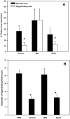
(A) Quantitative assessment of the extent of myonecrosis and regeneration in mouse gastrocnemius muscle injected with either B. asper venom, Mtx or BaP1. Histological sections stained with hematoxylin and eosin were analyzed one and 28 days after injections. The extent of necrosis was estimated in samples collected one day after injection as the percentage of the examined area corresponding to necrotic fibers, whereas the extent of regeneration was estimated in samples collected at 28 days as the percentage of the examined area corresponding to regenerating fibers, i.e. fibers having centrally-located nuclei. *p < 0.05 when comparing the percentage of necrosis and of regeneration for a single treatment. (B) Quantitative assessment of the diameter of regenerating muscle fibers, i.e. fibers presenting centrally-located nuclei, 28 days after intramuscular injection in the gastrocnemius of PBS, B. asper venom, Mtx or BaP1. Regenerating fibers in tissue injected with venom and BaP1 showed a reduced diameter when compared with control fibers in tissue injected with PBS (p < 0.05), whereas no significant difference was observed in the diameter of regenerating fibers in muscle injected with Mtx (p > 0.05). In both graphs, results are presented as mean±SD (n = 9).
Figure 3
Figure 3. Time-course of changes in capillary/muscle fiber ratio (A) and in capillary density (capillaries per mm2, B) in mouse gastrocnemius muscle injected with B. asper venom; control mice were injected with PBS.
Capillaries were visualized by immunohistochemistry with a rat anti-mouse CD31 monoclonal antibody, as described in Materials and Methods. A reduction in both parameters of capillary density occurred rapidly after venom injection, followed by a revascularization process. Results are presented as mean±SD (n = 9). * p < 0.05 when compared with capillary/muscle cell ratio and capillary density of muscle injected with PBS. (C–E) Immunohistochemical staining of endothelial cells in sections of mouse gastrocnemius muscle one day after injection of PBS (C), or one day (D) and seven days (E) after injection of B. asper venom. Endothelial cells were detected with anti-CD31 antibody, followed by a polyclonal biotinylated rabbit anti-rat IgG and Streptavidin Alexa fluor 488 (see Methods for details). Notice the loss of endothelial cell immunostaining in some areas in muscle injected with venom (asterisk). Bar represents 50 µm.
Figure 4
Figure 4. Apoptosis in regenerating skeletal muscle after the injection of either B. asper venom or Mtx.
Sections from muscle tissue samples collected 3, 5 and 7 days after injection of venom or Mtx were immunostained with anti-desmin antibodies and with TUNEL (see Methods for details). (A) The percentage of desmin-positive regenerating muscle fibers showing at least one TUNEL-positive nuclei, in relation to the total number of desmin-positive regenerating muscle fibers, was estimated. Results are presented as mean±SD (n = 9). (B) TUNEL-positive nuclei in desmin-positive cells 5 days after injection of either venom or Mtx. To highlight the pattern of spatial heterogeneity, each point corresponds to the density of TUNEL-positive nuclei per area in separate microscopic fields in different areas of the tissue. (C) and (D) Micrographs of muscle tissue sections from mice 5 days after injection of Mtx (C) or B. asper venom (D) immunostained for desmin (green fluorescence) and with TUNEL (reddish coloration in nuclei). No TUNEL-positive regenerating fibers, showing centrally-located nuclei, are observed in muscle injected with Mtx, whereas several regenerating fibers present TUNEL-positive nuclei in tissue injected with venom (arrows). Bar represents 50 µm.
Figure 5
Figure 5
(A) Changes in the density of intramuscular nerves in muscle tissue of mice 3 and 28 days after injection of either PBS, B. asper venom, Mtx or BaP1. Axons in nerves were visualized by immunostaining with a mouse anti-human neurofilament protein, as described in Materials and Methods. The number of intramuscular nerves per mm2 of tissue area showing at least one immunostained axon was quantified. Results are presented as mean±SD (n = 5). A significant drop (* p < 0.05) in nerves per area was observed at 3 days in samples injected with either venom, Mtx or BaP1, as compared to samples injected with PBS, whereas no differences in the number of nerves per area were detected between treatments at 28 days. (B) Axonal density in intramuscular nerves 28 days after injection of the various agents. The number of axons within each nerve was determined and expressed in terms of axons per nerve area. Results are presented as mean±SD (n = 11). * p < 0.05 when compared with axonal density in control muscles injected with PBS. **p < 0.05 when compared with axonal density in muscles injected with Mtx. (C to F) Light micrograph sections of mouse muscle tissue collected 28 days after injection of (C) PBS, (D) B. asper venom, (E) Mtx, and (F) BaP1. Sections were immunostained for neurofilament protein to detect axons in nerves (arrows). Notice the evident drop in the number of axons in samples from tissue injected with venom or BaP1. Bar represents 50 µm.

References

    1. Gutiérrez JM, Theakston RDG, Warrell DA. Confronting the neglected problem of snake bite envenoming: the need for a global partnership. PLoS Med. 2006;3:e150. - PMC - PubMed
    1. World Health Organization. A Neglected Public Health Issue. WHO, Geneva; 2007. Rabies and Envenomings.
    1. Kasturiratne A, Wickremasinghe AR, de Silva N, Gunawardena NK, Pathmeswaran A, et al. The global burden of snakebite: a literature analysis and modelling based on regional estimates of envenoming and deaths. PLoS Med. 2008;5:e218. - PMC - PubMed
    1. Fan HW, Cardoso JL. Clinical toxicology of snakebites in South America. Meier J, White J, editors. Handbook of Clinical Toxicology of Animal Venoms and Poisons. Boca Raton: CRC Press. 1995:667–688.
    1. Gutiérrez JM. Snakebite envenomation in Central America. Mackessy SP, editor. Handbook of Venoms and Toxins of Reptiles. Boca Raton: CRC Press. 2010:491–507.

Publication types