. 2011 Sep;60(9):1333-46.
doi: 10.1007/s00262-011-1030-6. Epub 2011 Jun 1.

A peptide epitope derived from the cancer testis antigen HOM-MEL-40/SSX2 capable of inducing CD4⁺ and CD8⁺ T-cell as well as B-cell responses

Affiliations

A peptide epitope derived from the cancer testis antigen HOM-MEL-40/SSX2 capable of inducing CD4⁺ and CD8⁺ T-cell as well as B-cell responses

Frank Neumann et al. Cancer Immunol Immunother. 2011 Sep.

Abstract

Background: Antigen-derived HLA class I-restricted peptides can generate specific CD8(+) T-cell responses in vivo and are therefore often used as vaccines for patients with cancer. However, only occasional objective clinical responses have been reported suggesting the necessity of CD4(+) T-cell help and possibly antibodies for the induction of an effective anti-tumor immunity in vivo. The SSX2 gene encodes the cancer testis antigen (CTA) HOM-MEL-40/SSX2, which is frequently expressed in a wide spectrum of cancers. Both humoral and cellular immune responses against SSX2 have been described making SSX2 an attractive candidate for vaccine trials.

Methods: SYFPEITHI algorithm was used to predict five pentadecamer peptides with a high binding probability for six selected HLA-DRB1 subtypes (*0101, *0301, *0401, *0701, *1101, *1501) which are prevalent in the Caucasian population.

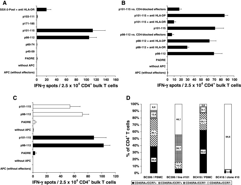
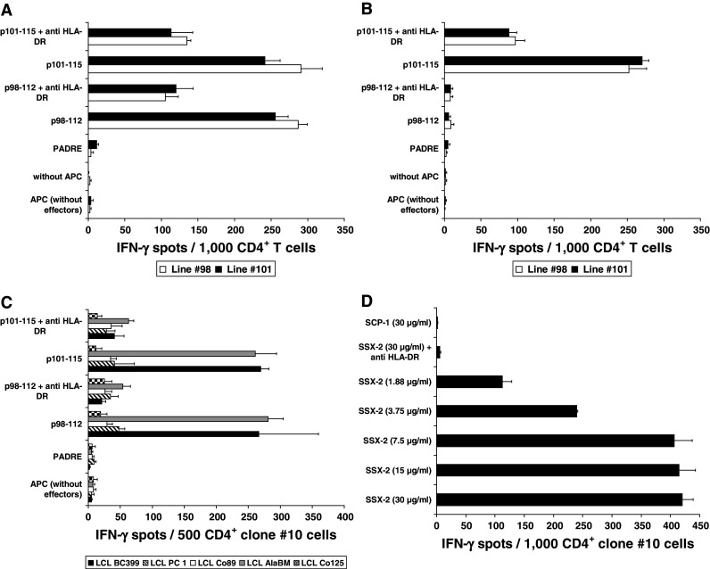
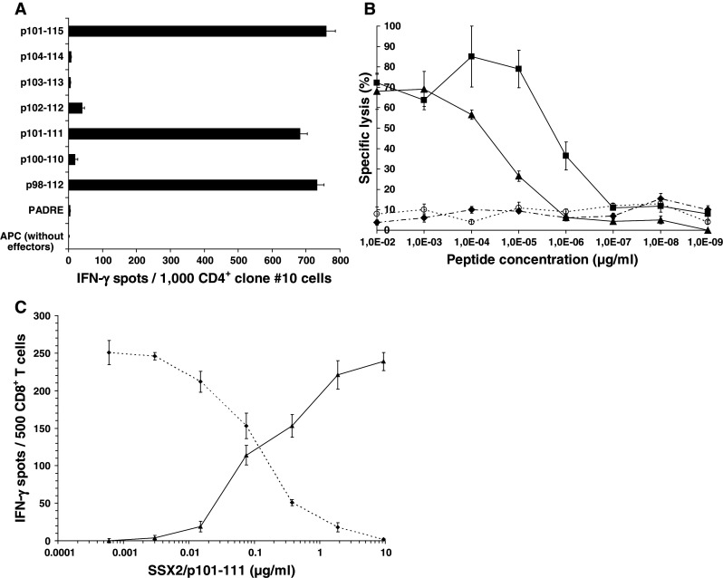
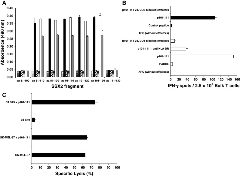
Results: Using peripheral blood cells of 13 cancer patients and 5 healthy controls, the HOM-MEL-40/SSX2-derived peptide p101-111 was identified as an epitope with dual immunogenicity for both CD4(+) helper and cytotoxic CD8(+) T cells. This epitope also reacted with anti-SSX2 antibodies in the serum of a patient with breast cancer. Most remarkably, SSX2/p101-111 simultaneously induced specific CD8, CD4, and antibody responses in vitro.

Conclusions: p101-111 is the first CTA-derived peptide which induces CD4(+), CD8(+), and B-cell responses in vitro. This triple-immunogenic peptide represents an attractive vaccine candidate for the induction of effective anti-tumor immunity.


Figures

Fig. 1
Fig. 1
CD4+ T-cell responses against HOM-MEL-40/SSX2-derived peptides and characterization of the CD4+ effector T cells. T cells from patients BC399 (a) and BC418 (b) responded after priming with the peptide pool to challenge with both p98-112 and p101-15. The response of T-cells from both patients against both peptides was blocked by anti-HLA-DR, but not by anti-HLA-DP. T cells from patient BC418 were also blocked by anti-CD4. c Response of bulk T cells from patient BC399 using single peptide p98-112 or p101-115. Filled bars represent the IFN-γ response of p98-112 primed bulk T cells when challenged with p98-112 (P = 0.003) and p101-115 (P < 0.02), respectively. Empty bars represent the response of T cells primed with p101-115 and challenged with p98-112 (P < 0.007) and p101-115 (P = 0.04), respectively. Y axes display the different APC settings used to challenge T cells during IFN-γ ELISPOT. d During stimulation with peptide p101-115 (patient BC399) and p98-112 (patient BC418), respectively, effector memory T cells of both patients increased considerably. CD4+ T cells of clone #10 all belong to the effector memory type
Fig. 2
Fig. 2
Dissection of the HLA-DR restriction of the T-cell response against SSX2-derived peptides p98-112 and p101-115 and natural processing and presentation of epitope p98-112. a Restriction of the T-cell response against p98-112 and p101-115 using the melanoma cell line SK-MEL-37 as APC. This cell line shares only DRB1*0101 with the BC399-derived T cells from line #98 and #101. DR restriction was demonstrated by blocking the response of both T-cell lines against APC pre-incubated with the anti-DR antibody clone L243. b DRB1*0701 restriction of the response from T cells of the BC399-lines #98 and #101 against LCL Co89 pulsed with peptide p101-115. *0701 is the only DRB1 subtype shared by Co89 and patient BC399. c Dissection of the restriction of the T-cell response of clone #10 from patient BC418: a response after stimulation with p98-112 or p101-115 only was observed when these peptides were presented by LCL from BC399 and AlaBM (P < 0.01) proving the restriction of these T cells to HLA-DRB1*0101, which is the only DR molecule shared by effector cells and APC. There was no response if both peptides were presented on LCL from PC 1, Co89, or Co125 excluding a restriction to HLA-DRB1*0301 or DRB3*0202. Y-axes of the A, B, and C panels display the different APC settings used to challenge T cells during IFN-γ ELISPOT. d Loading of autologous DC (1 × 104/well) with increasing concentrations of SSX2 whole protein induced a concentration-dependent response of clone #10 CD4+ T cells, which was blocked by anti-HLA-DR. The control antigen (SCP1 fragment p630-817) was not recognized (P < 0.001). Y axis shows the different DC settings used to challenge T cells during IFN-γ ELISPOT
Fig. 3
Fig. 3
Determination of peptide p101-111 as immunogenic core of the two peptides p98-112 and p101-115 and demonstration of the cytotoxic T-cell response against the core epitope p101-111. a After priming with peptide p98-112 CD4+ T cells of clone #10 (BC418) responded equally in IFN-γ ELISPOT when challenged with the pentadecamers p98-112 and p101-115 (P < 0.001) presented by allogeneic APC (3 × 104 LCL BC399/well). Peptide p101-111 was the only undecamer peptide to induce a response by clone #10 T cells (P < 0.001). Y axis displays the different APC settings used during IFN-γ ELISPOT. b HLA-A*0201-restricted CD8+ T cells from clone #24, which were generated by in vitro priming with the SSX2-derived nonamer p103-111, were used as effector cells in a CR51 release assay against HLA-A*0201+ T2 cells pulsed with decreasing concentrations of peptide p101-111 (triangles) or p103-111 (squares) by an E/T ratio of 10:1. Specific lysis of peptide-pulsed targets induced by clone #24 CD8+ T cells demonstrated a strong and dose-dependent cytotoxic T-cell response in an HLA-A*0201-restricted way. In contrast, the CTL epitope p41-49 (diamonds/dashed line), also derived from SSX2, as well as the DRB1*0101-restricted pentadecamer p98-112 (empty circles/spotted line) did not cause any cytotoxicity in the context of HLA-A*0201. c Further evidence for the HLA-A*0201 restriction of the CD8+ T cells after SSX2/p101-111 stimulation was shown by displacing an HLA-A*0201 reference peptide out of its MHC molecules, leading to modified antigen presenting functions of the APC and to a different induction pattern for T-cell responses finally. Increasing concentrations of the triple epitope resulted in a decreasing response of the HLA-A*0201-restricted IMP-specific clone #46 (diamonds/dashed line) and in a similarly increasing activity of the SSX2/p101-111 specific clone #9 (triangles/continuous line). Read out by ELISPOT against T2 cells pulsed with 0.1 μg/ml of the A*0201 reference peptide IMP/p58-66 either alone (1st concentration) or concurrently with increasing concentrations of SSX2/p101-111 (2nd-7th concentration). Both clones were run in separate experiments using T2 cells prepared in the same way
Fig. 4
Fig. 4
Simultaneous induction of a response by T-helper cells and by cytotoxic T cells as well as antibody production after in vitro stimulation with the SSX2-derived epitope p101-111. a After 35 days of in vitro stimulation of PBMC derived from patient BC699 with p101-111, the supernatant was analyzed by ELISA for antibodies binding to recombinantly expressed SSX2 fragments spanning the amino acids sequences as indicated on the X axis. This supernatant contained antibodies binding concentration dependent to SSX2 101-110 (empty [1:5-diluted] and gray [1:10-diluted] bars) and showed the same binding pattern (P < 0.01) as the positive serum derived from a melanoma cancer patient (black bars). At the beginning of the stimulation, such antibodies had not been present (horizontal stripes). A parallel stimulation using CMV-derived control peptides did not induce antibodies with affinity to any of the SSX2 fragments (striped bars [1:10]). SSX2 control serum also induced no significant ELISA signals (checkered bars). Only antibodies of the IgG class were analyzed. b PBMC from the HLA-A*0201, HLA-DRB1*0101 patient BC699 who had an SSX2-expressing tumor, were stimulated in vitro for 28 days with the epitope p101-111. The bulk lymphocytes developed simultaneously a strong T-cell response mediated by CD4+ T-helper cells as wells as by CD8+ cytotoxic T cells as shown by IFN-γ- release in the ELISPOT assay. Empty bars represent the response of 2.5 × 104 CD4+ bulk T cells/well against autologous PBMC (5 × 104/well) pulsed and labeled with the indicated peptide (4 μg/ml) and antibodies, respectively. Black bars represent the response of 2.5 × 104 CD8+ bulk T cells/well against T2 cells (3 × 104/well) pulsed and labeled with the indicated peptide (4 μg/ml) and antibodies, respectively. Y axis shows the different APC conformations. c After 35 days of in vitro stimulation with the undecamer peptide p101-111, the CD8+ T-cell fraction of the bulk culture of BC699 had developed a strong cytotoxic activity as assessed by LDH release assay. Lysis of cells of the HLA-A*0201+ and SSX2+ melanoma cell line SK-MEL-37 without external peptide pulsing demonstrated that epitope p101-111 is processed and presented via the MHC-I pathway. The HLA-A*0201+, but SSX2 line BT 549 caused lysis only after external pulsing with this peptide. Y axis displays the different APC conformations

Similar articles

Cited by

References

    1. Chen YT, Scanlan MJ, Sahin U, Tureci O, Gure AO, Tsang S, et al. A testicular antigen aberrantly expressed in human cancers detected by autologous antibody screening. Proc Natl Acad Sci USA. 1997;94:1914–1918. doi: 10.1073/pnas.94.5.1914. - DOI - PMC - PubMed
    1. Schultz ES, Lethe B, Cambiaso CL, Van Snick J, Chaux P, Corthals J, et al. A MAGE-A3 peptide presented by HLA-DP4 is recognized on tumor cells by CD4+ cytolytic T lymphocytes. Cancer Res. 2000;60:6272–6275. - PubMed
    1. Preuss KD, Zwick C, Bormann C, Neumann F, Pfreundschuh M. Analysis of the B-cell repertoire against antigens expressed by human neoplasms. Immunol Rev. 2002;188:43–50. doi: 10.1034/j.1600-065X.2002.18805.x. - DOI - PubMed
    1. Clark J, Rocques PJ, Crew AJ, Gill S, Shipley J, Chan AM, et al. Identification of novel genes, SYT and SSX, involved in the t(X;18)(p11.2;q11.2) translocation found in human synovial sarcoma. Nat Genet. 1994;7:502–508. doi: 10.1038/ng0894-502. - DOI - PubMed
    1. Tureci O, Chen YT, Sahin U, Gure AO, Zwick C, Villena C, et al. Expression of SSX genes in human tumors. Int J Cancer. 1998;77:19–23. doi: 10.1002/(SICI)1097-0215(19980703)77:1<19::AID-IJC4>3.0.CO;2-2. - DOI - PubMed

Publication types

MeSH terms