. 2011 Aug;41(8):2165-75.
doi: 10.1002/eji.201141728. Epub 2011 Jul 4.

The earliest intrathymic precursors of CD8α(+) thymic dendritic cells correspond to myeloid-type double-negative 1c cells

Affiliations

The earliest intrathymic precursors of CD8α(+) thymic dendritic cells correspond to myeloid-type double-negative 1c cells

Hervé Luche et al. Eur J Immunol. 2011 Aug.

Abstract

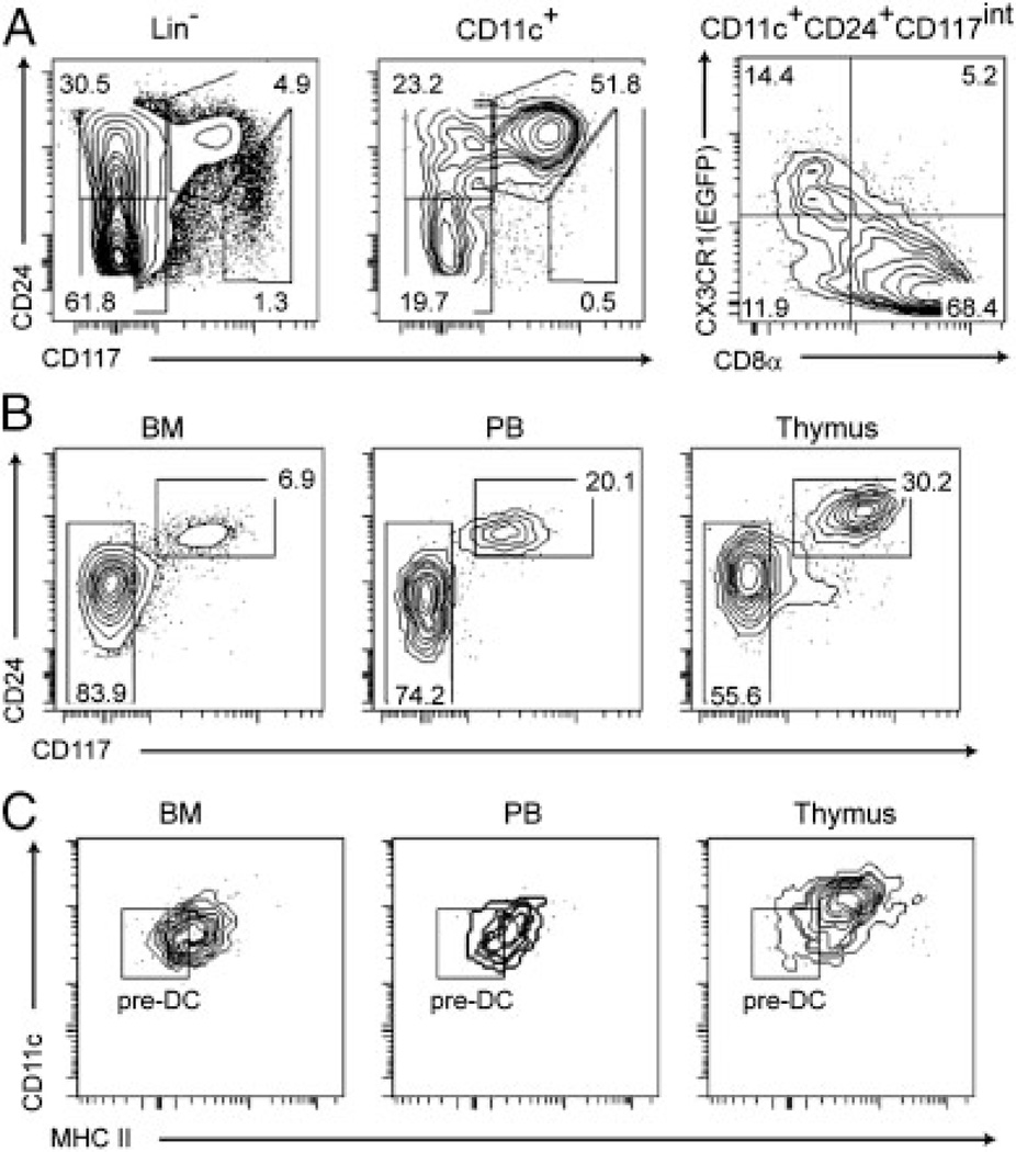
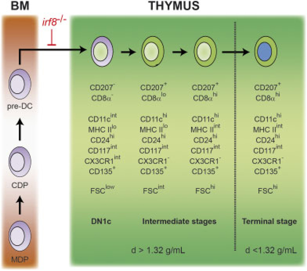
The dendritic cells (DCs) present in lymphoid and non-lymphoid organs are generated from progenitors with myeloid-restricted potential. However, in the thymus a major subset of DCs expressing CD8α and langerin (CD207) appears to stand apart from all other DCs in that it is thought to derive from progenitors with lymphoid potential. Using mice expressing a fluorescent reporter and a diphtheria toxin receptor under the control of the cd207 gene, we demonstrated that CD207(+) CD8α(+) thymic DCs do not share a common origin with T cells but originate from intrathymic precursors that express markers that are normally present on all (CD11c(+) and MHCII molecules) or on some (CD207, CD135, CD8α, CX3CR1) DC subsets. Those intrathymic myeloid-type precursors correspond to CD44(+) CD25(-) double-negative 1c (DN1c) cells and are continuously renewed from bone marrow-derived canonical DC precursors. In conclusion, our results demonstrate that the earliest intrathymic precursors of CD8α(+) thymic DCs correspond to myeloid-type DN1c cells and support the view that under physiological conditions myeloid-restricted progenitors generate the whole constellation of DCs present in the body including the thymus.


Conflict of interest statement

Conflict of interest: The authors declare no financial or commercial conflict of interest.

Figures

Figure 1
Figure 1
CD207+ tDCs correspond to CD8αhighCD172α−tDCs. Single-cell suspensions were prepared from thymi digested with collagenase-DNase I, and light-density cells were separated by centrifugation over an Optiprep gradient. After excluding eosinophils (CD11cintCD11bhigh), pDCs (CD11cintCD45R+) and NK cells (CD11cintNK1.1+), the remaining CD11cinttohiCD45RNK1.1 DCs were analyzed for the expression of CD207(EGFP) versus CD172α, CD8α versus CD172α and CD24 versus CD172α. Data are representative of at least ten independent experiments.
Figure 2
Figure 2
DN1c cells express markers that are normally present on DCs. Thymocytes of Langerin-EGFP or CX3CR1-EGFP mice were subjected to Optiprep gradient separation, and the heavy-density cell fraction was isolated and depleted of DP and of CD4+ SP T cells. Residual T cells, B cells and pDCs were excluded from further analysis using a cocktail of anti-CD3ε, anti-CD5 and anti-CD45R antibodies. (A) The remaining CD3εCD5CD45R DN cells were analyzed for the expression of CD44 versus CD25. CD11c+ cells constituted 0.5% of the DN cells and were exclusively found among the DN1 (CD44+CD25∼) subset. (B) DN1 cells were further subdivided into DN1a–b, DN1c, DN1d and DN1e subsets using CD24 and CD117 expression [41]. CD11c+ cells fell in the DN1c, DN1d and DN1e gates. (C) Expression of CD11c, CD207(EGFP), CD135, CD8α, MHCII and CX3CR1(EGFP) among the four DN1 subsets defined in (B). Percentages of cells positive for the specified marker are indicated. (D) Characterization of the DN1c subset of Langerin-EGFP mice. CD11c+ DN1 cells were separated into a CD207(EGFP)+ and a CD207(EGFP) fraction and analyzed for the expression of CD24 versus CD117. Gated CD207+ and CD207 DN1c cells were analyzed for the expression of CD11c, CD207(EGFP), CD135, CD8α, MHCII and for their size (FSC-A). The small fraction of CD135+ DN1a–b cells corresponded to the earliest thymus seeding progenitors [16, 17]. The DN1d and DN1e subsets contained small percentages of CD11c+CD207 cells that might correspond to CD207CD172α+ tDC precursors. Percentages of cells positive for the specified marker are indicated. The mean FSC-A value is indicated. Data are representative of three independent experiments.
Figure 3
Figure 3
Kinetics of reappearance of the CD11c+CD207+ cells found in the heavy-density cell fraction after DT treatment. (A) DN cells from the heavy-density cell fraction of Langerin-EGFP thymi were analyzed at various time points after the last DT injection for the expression of CD24 versus CD117 and CD11c versus CD117. Gates corresponding to the DN1a–b, DN1c, DN1d and DN1e subsets are as specified in Fig. 2B. (B) Gated CD11c+ DN1 cells were analyzed as in (A). (C) Gated CD11c+ DN1c cells were analyzed for CD207 versus CD8α expression at various time points after DT injection. Data are representative of three independent experiments and the percentages of cells found in each gate are indicated.
Figure 4
Figure 4
Kinetics of reconstitution of the CD207+ DCs found in the heavy-density and light-density cell fractions. (A) Kinetics of disappearance and reappearance of the CD207+ tDCs found in the heavy-density and light-density cell fractions of Langerin-DTREGFP thymi at various time points after the last DT injection. Absolute numbers of cells were determined using Flow count fluorospheres (Coulter) and normalized to the absolute numbers of cells present in thymi of Langerin-DTREGFP mice that received no DT. (B) BrdU was administered continuously for 12 days to a group of two to three B6 mice to compare the BrdU-labeling kinetics of the CD207+ tDCs found in the light-density and high-density fractions of an Optiprep gradient. Data in (A) are representative of at least three mice per time point and correspond to three independent experiments and data in (B) are representative of three to four independent experiments. Error bars correspond to the SEM.
Figure 5
Figure 5
IRF-8 is required for both DN1c and CD207+CD8αhigh tDC development. (A) DN cells from the heavy-density cell fraction of wild-type (WT) and irf8BHX2/BXH2 mice were analyzed for the expression of CD24 versus CD117. Gates corresponding to the DN1a–b, DN1c, DN1d and DN1e subsets are as specified in Fig. 2B. Numbers indicate the percentages of cells within the specified gates. (B) tDCs from the light-density cell fraction of wild-type (WT) and irf8BHX2/BXH2 mice were analyzed for the expression of CD24 versus CD172α. In (A), a cocktail of antibodies directed against CD3ε, CD25, CD45R, NK11 and CD11b permitted to exclude cells positive for those markers and to focus on ‘lineage negative’ (Lin) cells. Data in (A) and (B) are representative of four mice per genotype and correspond to two independent experiments. Numbers indicate the percentages of cells within the specified gates.
Figure 6
Figure 6
Cells resembling thymic DN1c cells can be found in the bone marrow and the blood. (A) Lin DN1 cells of CX3CR1-EGFP mice were prepared as described in Fig. 2. DN1 and CD11c+ DN1 cells were analyzed for CD24 and CD117 expression. The pattern of CX3CR1(EGFP) versus CD8α is shown for CD11c+ DN1c (CD24+CD117int) cells. (B) CD11c+ cells from bone marrow (BM) peripheral blood (PB) and thymus of CX3CR1-EGFP mice were enriched by MACS separation. After removing cells expressing CD25, CD45R, NK1.1, CD3e, CD19, Gr-1, CD115, CD172a, Sca-1 or CD8α, the remaining LinCX3CR1(EGFP)low cells were analyzed for the expression of CD24 versus CD117. (C) CD24+CD117int cells were prepared from bone marrow (BM) peripheral blood (PB) and thymus of CX3CR1-EGFP mice as defined in (B) and analyzed for CD11c versus MHCII molecules. Gate has been set up using the MHCIICD24lowCD117 cells shown in panel (B). Results are representative of two independent experiments.
Figure 7
Figure 7
Thymic and splenic CD8α+ DCs arise from MDPs, CDPs and pre-DCs. MDPs, CDPs and pre-DCs purified from the BM of CD45.2+ mice were adoptively transferred into unconditioned CD45.1+ congenic host. Thymus (A) and spleen (B) were analyzed a week after adoptive transfer. CD11c+ cells were enriched using magnetic beads coated with anti-CD11c antibody and CD11c+MHCII+ DCs were identified on a CD11c versus MHCII dot plot. CD11c+MHCII+ DCs were subsequently analyzed and separated into CD8αhigh and CD8αlow fractions. Percentages of host-derived (CD45.1+) and donor-derived (CD45.2+) cells are shown for the CD11c+CD8αhigh and CD11c+CD8αlow fractions. Results are representative of three independent experiments.
Figure 8
Figure 8
A model of CD207+CD8αhigh tDC development. Common macrophage-DC progenitors (MDP), common DC precursor (CDP) and classical DC-restricted precursors (pre-DCs) present in the bone marrow (BM) generate CD207+CD8αhigh tDCs. The earliest stage of the intrathymic developmental series corresponds to the DN1c (CD11c+ CD207CD8αMHCIIlow) cells previously identified by Porritt and colleagues [14]. The DN1c cells are found in the heavy-density cell fraction (d41.32g/mL) of an Optiprep gradient and progress through CD11c+CD207+CD8α and CD11c+CD207+CD8α+ intermediate stages to the mature CD207+CD8αhigh stage that is found in the light-density cell fraction (d< 1.32 g/mL) of an Optiprep gradient. The developmental block observed in mice expressing a mutation in the interferon-regulatory factor 8 (irf8) gene is shown. Our findings do not formally rule out the possibility that, under some experimental conditions, early T-cell precursors can give rise to CD207+CD8αhigh tDCs. As recently stressed [42], it is important to distinguish physiological fate choices - as documented in the present study - from cell fates that are possible experimentally.

Comment in

References

    1. Petrie HT, Zuniga-Pflucker JC. Zoned out: functional mapping of stromal signaling microenvironments in the thymus. Annu. Rev. Immunol. 2007;25:649–679. - PubMed
    1. McCaughtry TM, Baldwin TA, Wilken MS, Hogquist KA. Clonal deletion of thymocytes can occur in the cortex with no involvement of the medulla. J. Exp. Med. 2008;205:2575–2584. - PMC - PubMed
    1. Klein L, Hinterberger M, Wirnsberger G, Kyewski B. Antigen presentation in the thymus for positive selection and central tolerance induction. Nat. Rev. Immunol. 2009;9:833–844. - PubMed
    1. Gray D, Abramson J, Benoist C, Mathis D. Proliferative arrest and rapid turnover of thymic epithelial cells expressing Aire. J. Exp. Med. 2007;204:2521–2528. - PMC - PubMed
    1. Koble C, Kyewski B. The thymic medulla: a unique microenvironment for intercellular self-antigen transfer. J. Exp. Med. 2009;206:1505–1513. - PMC - PubMed

Publication types

MeSH terms

LinkOut - more resources