. 2011 Sep 1;98(3):372-82.
doi: 10.1002/jbm.a.33112. Epub 2011 May 31.

Vascular endothelial growth factor and fibroblast growth factor 2 delivery from spinal cord bridges to enhance angiogenesis following injury

Affiliations

Vascular endothelial growth factor and fibroblast growth factor 2 delivery from spinal cord bridges to enhance angiogenesis following injury

Laura De Laporte et al. J Biomed Mater Res A. .

Abstract

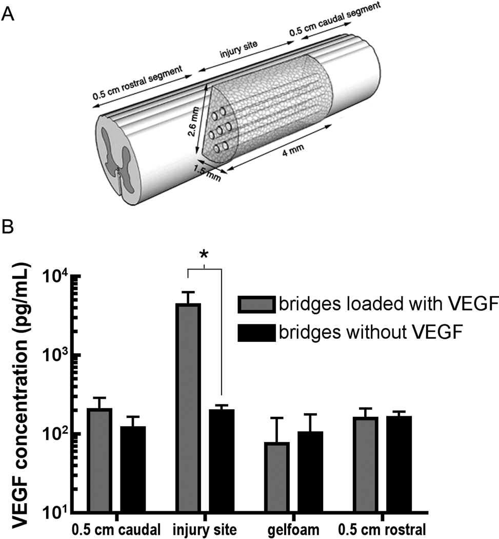
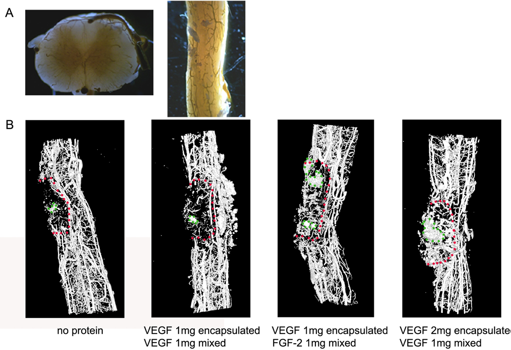
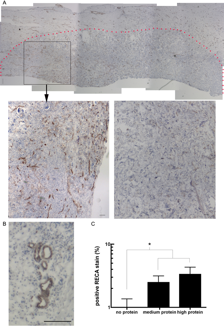
The host response to spinal cord injury can lead to an ischemic environment that can induce cell death and limits cell transplantation approaches to promote spinal cord regeneration. Spinal cord bridges that provide a localized and sustained release of vascular endothelial growth factor (VEGF) and fibroblast growth factor 2 (FGF-2) were investigated for their ability to promote angiogenesis and nerve growth within the injury. Bridges were fabricated by fusion of poly(lactide-co-glycolide) microspheres using a gas foaming/particulate leaching technique, and proteins were incorporated by encapsulation into the microspheres and/or mixing with the microspheres before foaming. Compared to the mixing method, encapsulation reduced the losses during leaching and had a slower protein release, while VEGF was released more rapidly than FGF-2. In vivo implantation of bridges loaded with VEGF enhanced the levels of VEGF within the injury at 1 week, and bridges releasing VEGF and FGF-2 increased the infiltration of endothelial cells and the formation of blood vessel at 6 weeks postimplantation. Additionally, substantial neurofilament staining was observed within the bridge; however, no significant difference was observed between bridges with or without protein. Bridges releasing angiogenic factors may provide an approach to overcome an ischemic environment that limits regeneration and cell transplantation-based approaches.


Figures

Figure 1
Figure 1
Protein encapsulation efficiency and protein retention after leaching. A) VEGF and FGF-2 encapsulation efficiency after microsphere encapsulation and collection. B) VEGF and FGF-2 retention after leaching bridges for 1 h. Proteins were incorporated using either the encapsulation only or mixing only approaches. Significant differences between protein conditions are denoted by an asterisk (p<0.05).
Figure 2
Figure 2
Protein release from bridge. Bridges were fabricated with VEGF (filled line) or FGF-2 (dotted line) in three different manners: mixing only (squares), encapsulation only (triangles), or a combination of both encapsulation and mixing (circles). The amount of protein left in the bridge after leaching was set as 100%.
Figure 3
Figure 3
VEGF and FGF-2 activity during in vitro release. Phosphorylation of (A) VEGF and (B) FGF-2 receptors was assessed by western blot. Legend: 1. control (100 ng/ml VEGF or 50 ng/ml FGF-2), 2. mixing 24 hours, 3. encapsulation 24 hours, 4. mixing 5 days, 5. encapsulation 5 days, 6. mixing 10 days, 7. encapsulation 10 days, 8. mixing 20 days, 9. encapsulation 20 days, 10. mixing 42 days, 11. encapsulation 42 days, 12. negative control (PBS).
Figure 4
Figure 4
ELISA for VEGF at the injury and in adjacent tissue at 1 week post implantation. A) Schematic of the spinal cord injury and segments that were analyzed for ELISA, adapted from De Laporte L, Yang Y, Zelivyanskaya ML, Cummings BJ, Anderson AJ, Shea LD. Plasmid releasing multiple channel bridges for transgene expression after spinal cord injury. Originally published in Molecular Therapy 2009;17(2):318–26 [6], with permission of the Nature Publishing Group. B) VEGF levels in different segments for bridges loaded with and without VEGF. Significant differences between conditions are denoted by an asterisk (p<0.05).
Figure 5
Figure 5
A) Cured Microfil injection compound that formed a three-dimensional cast of the rat’s spinal cord vasculature. From left to right: cross section and longitudinal view. B) 3D reconstructions of micro-CT scans of bridge implants in spinal cord hemisection model at 6 weeks post implantation. From left to right: bridge implant without protein loading, bridge implant with 1 µg VEGF encapsulated and 1 µg VEGF mixed, bridge implant with 1 µg VEGF encapsulated and 1 µg FGF-2 mixed, bridge implant with 2 µg VEGF encapsulated and 1 µg VEGF mixed. The red dotted line marks the contours of the bridge at the implant site. The green dotted line marks the contour of some residual Gelfoam that appeared on microCT. The Gelfoam was used to cover the injury site, and mostly removed upon tissue retrieval.
Figure 6
Figure 6
Endothelial cell infiltration in bridge at 6 weeks post implantation. A) RECA stain (brown): from left to right: bridge containing 4 µg of VEGF encapsulated within microspheres and 2 µg of FGF-2 and VEGF mixed (high protein), and bridge without protein. The red dotted line marks the contours of the bridge at the implant site. Scale bar: 200 µm. B) Blood vessels adjacent to the bridge containing 4 µg of VEGF encapsulated within microspheres and 1 µg of FGF-2 and VEGF mixed (medium protein). Scale bar: 200 µm. C) Quantifcation of RECA staining for the three conditions.
Figure 7
Figure 7
Neurite ingrowth with bridge at 6 weeks post implantation. A) Neurofilament stain (brown) of bridge implanted with the highest VEGF/FGF-2 protein dose. The red dotted line marks the contours of the bridge at the implant site. Scale bar: 500 µm. B) Quantification of positive NF stain for bridge implants without VEGF/FGF-2, and bridge implants with a medium and high dose of VEGF/FGF-2 protein.

References

    1. Storkebaum E, Lambrechts D, Carmeliet P. VEGF: once regarded as a specific angiogenic factor, now implicated in neuroprotection. Bioessays. 2004;26(9):943–954. - PubMed
    1. Jimenez Hamann MC, Tator CH, Shoichet MS. Injectable intrathecal delivery system for localized administration of EGF and FGF-2 to the injured rat spinal cord. Exp. Neurol. 2005;194(1):106–119. - PubMed
    1. Jones LL, Oudega M, Bunge MB, Tuszynski MH. Neurotrophic factors, cellular bridges and gene therapy for spinal cord injury. J Physiol. 2001;533(Pt 1):83–89. - PMC - PubMed
    1. Nakahara Y, Gage FH, Tuszynski MH. Grafts of fibroblasts genetically modified to secrete NGF, BDNF, NT-3, or basic FGF elicit differential responses in the adult spinal cord. Cell Transplant. 1996;5(2):191–204. - PubMed
    1. De Laporte L, Yan AL, Shea LD. Local gene delivery from ECM-coated poly(lactide-coglycolide) multiple channel bridges after spinal cord injury. Biomaterials. 2009;30(12):2361–2368. - PMC - PubMed

Publication types

MeSH terms