. 2011 Jun 1:8:61.
doi: 10.1186/1742-2094-8-61.

A dual role for microglia in promoting tissue inhibitor of metalloproteinase (TIMP) expression in glial cells in response to neuroinflammatory stimuli

Affiliations

A dual role for microglia in promoting tissue inhibitor of metalloproteinase (TIMP) expression in glial cells in response to neuroinflammatory stimuli

Jennifer V Welser-Alves et al. J Neuroinflammation. .

Abstract

Background: By neutralizing the effect of the matrix metalloproteinases (MMPs), the tissue inhibitors of matrix metalloproteinases (TIMPs) play a critical role in maintaining tissue proteolysis in balance. As the major reactive glial cell types in the central nervous system (CNS), microglia and astrocytes play fundamental roles in mediating tissue breakdown and repair. As such, it is important to define the TIMP expression profile in these cells, as well as the mechanisms of regulation by neuroinflammatory stimuli.

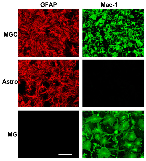
Methods: Primary mixed glial cultures (MGC), pure microglia, and pure astrocytes were used in this study. To study astrocytes, we employed a recently described pure astrocyte culture system, which has the major advantage of totally lacking microglia. The three different types of culture were treated with lipopolysaccharide (LPS) or individual cytokines, and cell culture supernatants assayed for TIMP-1 or TIMP-2 protein expression by western blot.

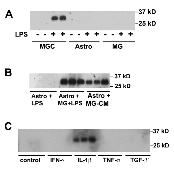
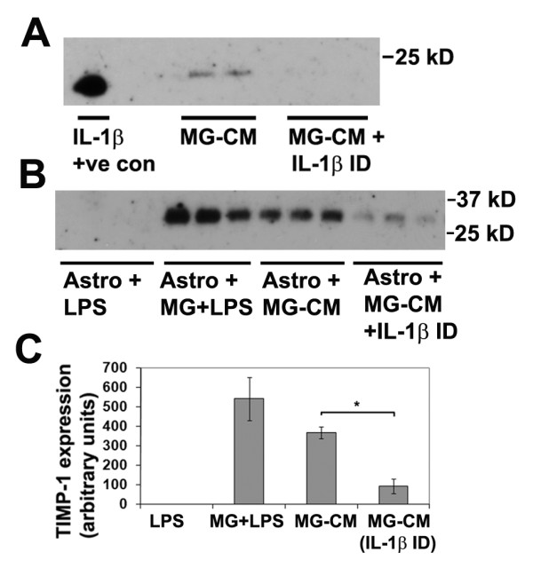
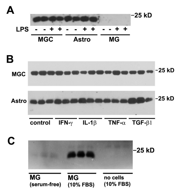
Results: LPS induced TIMP-1 expression in MGC, but not in pure astrocyte or microglial cultures. When pure astrocytes were treated with the cytokines IL-1β, IFN-γ, TNF or TGF-β1, only IL-1β induced TIMP-1 expression. Significantly, astrocyte TIMP-1 expression was restored in LPS-treated astrocyte cultures after the addition of microglia, or conditioned medium taken from LPS-activated microglia (MG-CM). Furthermore, this effect was lost after depletion of IL-1β from MG-CM. By contrast, TIMP-2 was constitutively expressed by astrocytes, whereas microglia expressed TIMP-2 only after exposure to serum.

Conclusions: Taken together, these results demonstrate an important concept in glial interactions, by showing that microglia play a central role in regulating glial cell expression of TIMPs, and identify microglial IL-1β as playing a key role in mediating microglial-astrocyte communication.


Figures

Figure 1
Figure 1
Defining the cell purity of the three different glial cell culture systems. Mixed glial cultures (MGC), pure astrocytes (Astro) and pure microglial cultures (MG) were established as described in Materials and Methods, and then analyzed by immunofluorescence for expression of the astrocyte marker GFAP (Cy3, red) or the microglial marker Mac-1 (AlexaFluor 488, green). Scale bar = 50 μm. Note that MGC contain both astrocytes and microglia. In contrast, neurosphere-derived pure astrocyte cultures contain only astrocytes with no microglia, whereas pure microglial cultures contain only microglia.
Figure 2
Figure 2
Analysis of glial cell TIMP-1 expression in response to cytokines and LPS. TIMP-1 expression was examined by western blot of cell culture supernatants, as described in Materials and Methods. A. The influence of LPS on TIMP-1 expression in the three different types of glial culture. MGC, astrocytes, and microglial cultures were treated with LPS for 2 days, then cell culture supernatants examined for TIMP-1 expression. Note that cultures not treated with LPS did not express TIMP-1, and TIMP-1 protein (~28 kD band) was induced only in MGC following LPS stimulation. B. TIMP-1 induction in astrocytes in the presence of activated microglia or MG-CM. Pure astrocyte cultures received either: LPS, microglia + LPS, or LPS-activated MG-CM. Note that the presence of LPS-activated microglia or LPS-activated MG-CM strongly induced TIMP-1 expression in astrocytes. C. The influence of cytokines on TIMP-1 expression by astrocytes. Pure astrocyte cultures were treated with a panel of different cytokines for 2 days, and the supernatants analyzed for TIMP-1 expression. Note that of the cytokines tested, only IL-1β stimulated TIMP-1 expression in astrocytes.
Figure 3
Figure 3
Examining the role of microglia-derived IL-1β in promoting astrocyte TIMP-1 expression. A. Determination of IL-1β in LPS-activated MG-CM. Microglial cultures were treated with LPS for 2 days, then subject to either six rounds of IL-1β immunodepletion, or mock depletion, as described in Materials and Methods, and samples analyzed for the presence of IL-1β. Note that IL-1β was detected in the LPS-activated MG-CM before, but not after the IL-1β immunodepletion, demonstrating the effectiveness of the immunodepletion strategy. B and C. The effect of removing IL-1β from LPS-activated MG-CM on astrocyte TIMP-1 production. Pure astrocyte cultures received either: LPS, microglia + LPS, LPS-activated MG-CM (mock depleted) or LPS-activated MG-CM depleted of IL-1β. Note that the presence of LPS-activated microglia or LPS-activated MG-CM strongly induced TIMP-1 expression in astrocytes, but this effect was markedly attenuated after IL-1β removal from the MG-CM. * p < 0.02.
Figure 4
Figure 4
Analysis of glial cell TIMP-2 expression in response to cytokines and LPS. TIMP-2 expression was examined by western blot of cell culture supernatants, as described in Materials and Methods. A. The influence of LPS on TIMP-1 expression in the three different types of glial culture. MGC, astrocytes, and microglial cultures were treated with LPS for 2 days, then cell culture supernatants examined for TIMP-2 expression. Note that MGC and pure astrocytes constitutively expressed TIMP-2 (~22 kD) band, and this expression was not enhanced by LPS stimulation. In contrast, microglial cultures did not express TIMP-2. B. The influence of cytokines on TIMP-2 expression by MGC and pure astrocytes. MGC and pure astrocyte cultures were treated with a panel of different cytokines for 2 days, and the supernatants analyzed for TIMP-1 expression. Note that both MGC and pure astrocytes constitutively expressed TIMP-2, and this expression level was not significantly affected by any of the cytokines tested. C. The influence of serum on microglial TIMP-2 expression. Pure microglial cultures were grown either in serum-free medium or in medium containing 10% FBS. After 2 days, the cell culture supernatants were analyzed for TIMP-2 expression. Note that the presence of serum strongly induced TIMP-2 expression in microglial cells.

References

    1. Crocker SJ, Pagenstecher A, Campbell IL. The TIMPs tango with MMPs and more in the central nervous system. J Neurosci Res. 2004;75:1–11. doi: 10.1002/jnr.10836. - DOI - PubMed
    1. Gardner J, Ghorpade A. Tissue inhibitor of metalloproteinase (TIMP)-1: the TIMPed balance of matrix metalloproteinases in the central nervous system. J Neurosci Res. 2003;74:801–806. doi: 10.1002/jnr.10835. - DOI - PMC - PubMed
    1. Agrawal SM, Lau L, Yong VW. MMPs in the central nervous system: where the good guys go bad. Semin Cell Dev Biol. 2008;19:42–51. doi: 10.1016/j.semcdb.2007.06.003. - DOI - PubMed
    1. Stetler-Stevenson WG. Tissue inhibitors of metalloproteinases in cell signaling: metalloproteinases-independent biological activities. Sci Signal. 2008;1:re6. doi: 10.1126/scisignal.127re6. - DOI - PMC - PubMed
    1. Candelario-Jalil E, Yang Y, Rosenberg GA. Diverse roles of matrixmetalloproteinsases and tissue inhibitors of metalloproteinases in neuroinflammation and cerebral ischemia. Neuroscience. 2009;158:983–994. doi: 10.1016/j.neuroscience.2008.06.025. - DOI - PMC - PubMed

Publication types

MeSH terms