

Review
doi: 10.1074/jbc.R111.225854.
Epub 2011 Jun 1.
Imaging of intact tissue sections: moving beyond the microscope
Affiliations
- PMID: 21632549
- PMCID: PMC3138310
- DOI: 10.1074/jbc.R111.225854
Item in Clipboard
Review
Imaging of intact tissue sections: moving beyond the microscope
J Biol Chem.
.
Abstract
MALDI-imaging MS is a new molecular imaging technology for direct in situ analysis of thin tissue sections. Multiple analytes can be monitored simultaneously without prior knowledge of their identities and without the need for target-specific reagents such as antibodies. Imaging MS provides important insights into biological processes because the native distributions of molecules are minimally disturbed, and histological features remain intact throughout the analysis. A wide variety of molecules can be imaged, including proteins, peptides, lipids, drugs, and metabolites. Several specific examples are presented to highlight the utility of the technology.
Figures
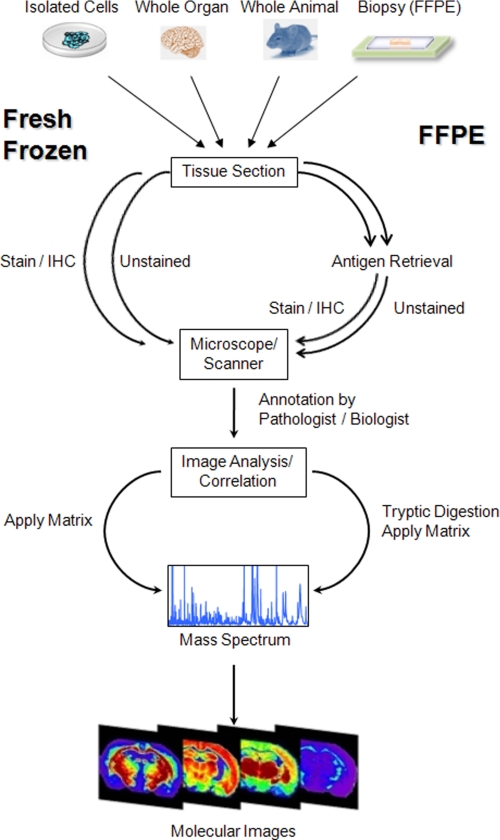
Schematic outline of a typical workflow for tissue samples. Sample pretreatment steps include cutting and mounting the tissue section on a conductive target as well as paraffin removal and antigen retrieval for FFPE sections. A frozen serial section or the FFPE section to be analyzed is stained, a digital image is taken, and areas of interest are selected by a pathologist. Trypsin and/or matrix is applied to the tissue section, and mass spectra are generated at each x,y coordinate. IHC, immunohistochemistry.
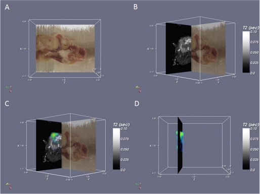
Three-dimensional IMS and co-registration. A, block face imaging of a rat head. Sections were collected axially but can be viewed in any orientation such as the sagittal view shown here. B, block face image (sagittal) co-registered with the T2 magnetic resonance image (axial). Brain structure, including the corpus callosum and a tumor, can be observed in the MRI data. C, MS data for a tumor-specific protein, astrocytic phosphoprotein PEA, are superimposed on the MRI data. D, three-dimensional rendered volume of the MS data (sagittal) on a single magnetic resonance plane (axial).
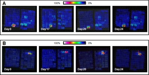
Reproducibility of on-tissue tryptic digestion from a TMA comprising samples from multiple different organ sites and cancers. A, a feature at m/z 1650.0 was found only in the leiomyosarcoma samples. B, a feature at m/z 1177.7 was present only in the astrocytoma samples. Both features were reproducible when comparing serial sections that were analyzed on 4 different days.
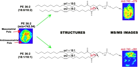
MS/MS lipid imaging. MS imaging of m/z 742.54 shows a ubiquitous pattern from top to bottom of the implantation site. However, MS/MS imaging of this ion shows that it is made up of two distinct isobaric structures that have very different localization patterns. PE (36:2; 18:0/18:2) is localized to the antimesometrial pole (lower), whereas PE (36:2; 18:1/18:1) is localized to the mesometrial pole (upper).
Comment in
-
Thematic minireview series on biological applications of mass spectrometry.J Biol Chem. 2011 Jul 22;286(29):25417. doi: 10.1074/jbc.R111.266700. Epub 2011 Jun 1. J Biol Chem. 2011. PMID: 21632545 Free PMC article.
References
-
- Aerni H. R., Cornett D. S., Caprioli R. M. (2006) Anal. Chem. 78, 827–834 - PubMed
Publication types
MeSH terms
Substances
Grants and funding
LinkOut - more resources
Full Text Sources
