. 2012 Jan 22;279(1727):224-9.
doi: 10.1098/rspb.2011.0740. Epub 2011 Jun 1.

The presence of an embryonic opercular flap in amniotes

Affiliations

The presence of an embryonic opercular flap in amniotes

Jo Richardson et al. Proc Biol Sci. .

Abstract

The operculum is a large flap consisting of several flat bones found on the side of the head of bony fish. During development, the opercular bones form within the second pharyngeal arch, which expands posteriorly and comes to cover the gill-bearing arches. With the evolution of the tetrapods and the assumption of a terrestrial lifestyle, it was believed that the operculum was lost. Here, we demonstrate that an embryonic operculum persists in amniotes and that its early development is homologous with that of teleosts. As in zebrafish, the second pharyngeal arch of the chick embryo grows disproportionately and comes to cover the posterior arches. We show that the developing second pharyngeal arch in both chick and zebrafish embryos express orthologous genes and require shh signalling for caudal expansion. In amniotes, however, the caudal edge of the expanded second arch fuses to the surface of the neck. We have detailed how this process occurs and also demonstrated a requirement for thyroid signalling here. Our results thus demonstrate the persistence of an embryonic opercular flap in amniotes, that its fusion mirrors aspects of amphibian metamorphosis and gives insights into the origin of branchial cleft anomalies in humans.


Figures
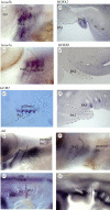
Figure 1.
Figure 1.
The second arches of fish and amniotes are homologous. (a) hoxa2b expression in the outgrowing opercular flap in a 3 dpf zebrafish embryo. (b) HOXA2 expression in PA2 of an E5 chick embryo. (c) hoxa3a is expressed in the developing gill arches (PA3+) but not in the opercular flap covering them in a 3 dpf zebrafish embryo. (d) HOXB3 expression in an E5 chick embryo, longitudinal section. The non-expressing second arch covers HOXB3+ve posterior pharyngeal region. (e) Longitudinal section through a 4 dpf zebrafish embryo showing gcm2 expression in the developing gill buds and inner edge of the expanding opercular flap. (f) Longitudinal section of an E5 chick embryo showing GCM2 expression in pouches 3 and 4, with the non-expressing expanding second arch. (g) Expression of shh at the caudal edge of the second arch in a 53 hpf zebrafish embryo. (h) Expression of shh at the leading edge of the expanding second arch of a 4 dpf zebrafish embryo, (i) SHH expression in an E3 (HH18) chick embryo at the posterior edge of the growing second arch. (j) Pronounced SHH expression at the caudal edge of the growing PA2 in an E4 (HH23) chick embryo. The edge of the second arch is outlined in (a–f).
Figure 2.
Figure 2.
Shh inhibition reduces the outgrowth of the second arch in chick and zebrafish. (a,b) Five dpf zebrafish embryos treated with ethanol (carrier, a), or cyclopamine (b), and probed with shh to visualize the flap. Numbers indicate the measurements taken to calculate the size of the flap. (c,d) Bissected chick embryos (4 dpf) treated with (c) HBC—carrier or (d) cyclopamine. The second arch (PA2) and otic vesicle (OV) are outlined in red. (e) Graph showing the size of the opercular flap (OF) in treated fish as a percentage of total eye–pectoral fin length. (f) Graph showing the area of the second arch (PA2) and otic vesicle (OV; internal control to account for any generalized growth reduction) in squared micrometre. There is a significant reduction in PA2 compared with the OV for the cyclopamine-treated embryos versus HBC (carrier) control-treated embryos (two-tailed t-test, p < 0.001 for both d and e). Blue, 15 µl HBC control; pink, 15 µl cyclopamine.
Figure 3.
Figure 3.
Morphogenesis of the amniote second arch and inhibition of fusion. (a) Longitudinal section of ectoderm labelled with CCFSE. The inner surface of the second arch is endodermally derived. The leading edge of the second arch is indicated by the white arrow. (b) Laminin immunofluoresence at E4; the second arch has not fused. Leading edge indicated by white arrow. (c) Laminin staining shows the beginning of fusion of the inner surface of the second arch to the subjacent epithelium of the poster arches at E5 (white arrow). Posterior edge of the second arch indicated by the white arrow. (d) Longitudinal section at E6 shows the second arch has broadly fused; internally, a small gap remains where the sinus is in the process of closing up (white asterisk). (e) These internal surfaces eventually fuse; the line of fusion marked by a white asterisk. (f) Lysotracker staining labelling apoptotic cells. Some dying cells are present along the line of fusion. (g) Treatment with thyroid-blocking drugs (amiodarone and methimazole) leads to failure of fusion (white arrowhead) and persistence of cyst (white asterisk). Longitudinal section at E6 (HH29). (h) A partial failure of fusion after treatment with thyroid hormone-blocking drugs (methimazole only). There is a persistent cyst (white asterisk) and an involuted structure where the ectodermal interfaces have failed to properly meet and fuse (white arrowhead). Longitudinal section at E6 (HH29). (i) Whole-mount chick embryo (E7) stained with lysotracker, showing a line of cell death at the remaining edge of the fused second arch, expanding medially from the lateral edges. ecto, ectoderm; endo, endoderm; PA2, pharyngeal arch 2.

References

    1. Brazeau M. D., Ahlberg P. E. 2006. Tetrapod-like middle ear architecture in a Devonian fish. Nature 439, 318–32110.1038/nature04196 (doi:10.1038/nature04196) - DOI - DOI - PubMed
    1. Daeschler E. B., Shubin N. H., Jenkins F. A., Jr 2006. A Devonian tetrapod-like fish and the evolution of the tetrapod body plan. Nature 440, 757–76310.1038/nature04639 (doi:10.1038/nature04639) - DOI - DOI - PubMed
    1. Kardong K. 1998. Vertebrates: comparative anatomy, function and evolution. New York, NY: McGraw-Hill
    1. Okabe M., Graham A. 2004. The origin of the parathyroid gland. Proc. Natl Acad. Sci. USA 101, 17 716–17 71910.1073/pnas.0406116101 (doi:10.1073/pnas.0406116101) - DOI - DOI - PMC - PubMed
    1. Schoenwolf G. C., Bleyl S. B., Brauer P. R., Francis-West P. H. 2009. Larsen's human embryology. New York, NY: Churchill Livingstone

Substances

LinkOut - more resources