. 2011 Sep;168(9):921-9.
doi: 10.1176/appi.ajp.2011.11010052. Epub 2011 Jun 1.

Cortical deficits of glutamic acid decarboxylase 67 expression in schizophrenia: clinical, protein, and cell type-specific features

Affiliations

Cortical deficits of glutamic acid decarboxylase 67 expression in schizophrenia: clinical, protein, and cell type-specific features

Allison A Curley et al. Am J Psychiatry. 2011 Sep.

Abstract

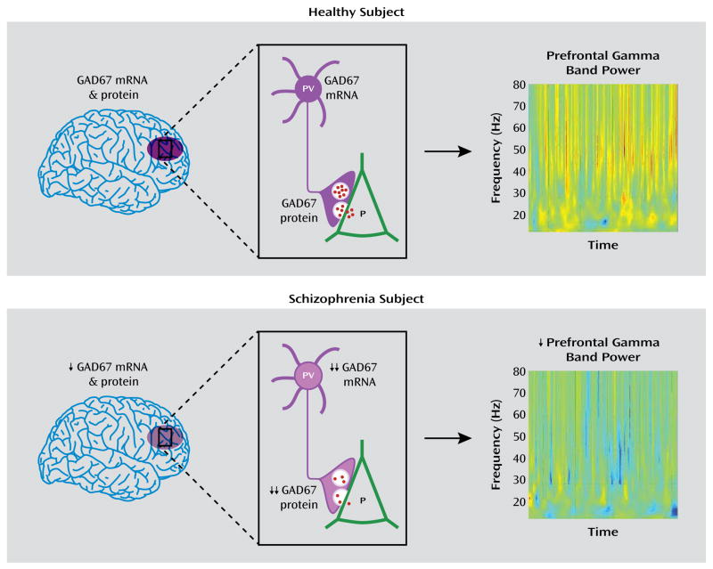
Objective: Cognitive deficits in schizophrenia are associated with altered activity of the dorsolateral prefrontal cortex, which has been attributed to lower expression of the 67 kDa isoform of glutamic acid decarboxylase (GAD67), the major γ-aminobutyric acid (GABA)-synthesizing enzyme. However, little is known about the relationship of prefrontal GAD67 mRNA levels and illness severity, translation of the transcript into protein, and protein levels in axon terminals, the key site of GABA production and function.

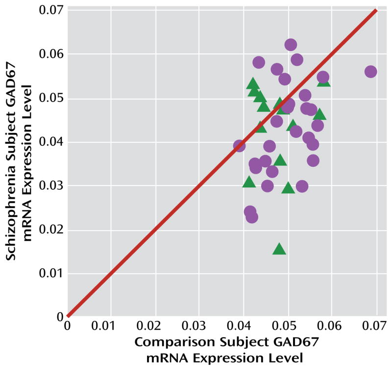
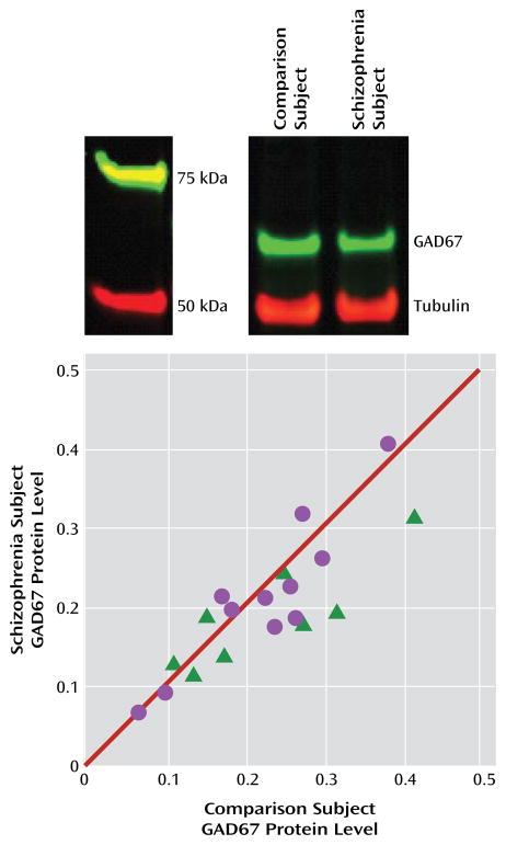
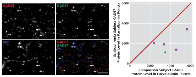
Method: Quantitative polymerase chain reaction was used to measure GAD67 mRNA levels in postmortem specimens of dorsolateral prefrontal cortex from subjects with schizophrenia and matched comparison subjects with no known history of psychiatric or neurological disorders (N=42 pairs). In a subset of this cohort in which potential confounds of protein measures were controlled (N=19 pairs), Western blotting was used to quantify tissue levels of GAD67 protein in tissue. In five of these pairs, multilabel confocal immunofluorescence was used to quantify GAD67 protein levels in the axon terminals of parvalbumin-containing GABA neurons, which are known to have low levels of GAD67 mRNA in schizophrenia.

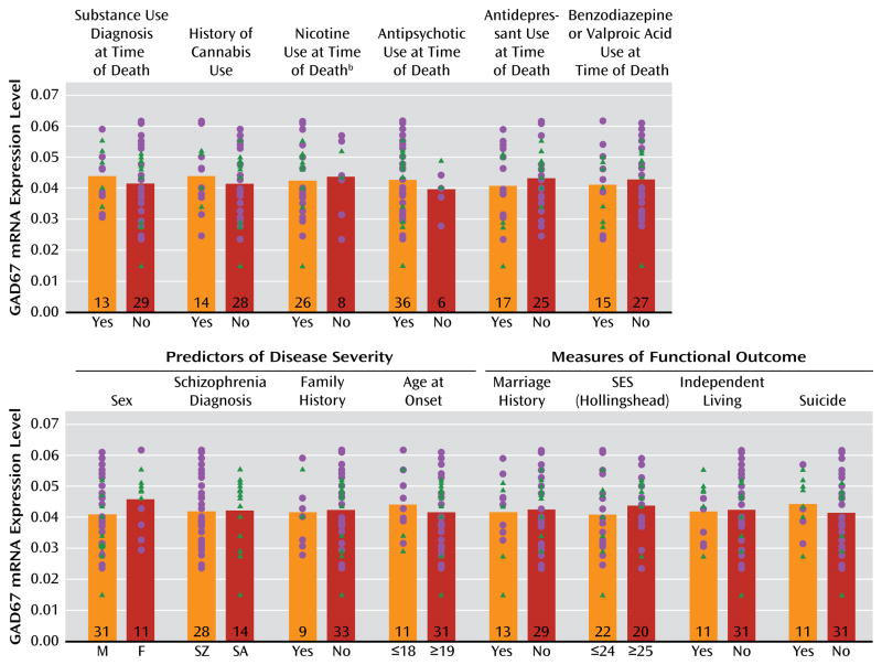
Results: GAD67 mRNA levels were significantly lower in schizophrenia subjects (by 15%), but transcript levels were not associated with predictors or measures of illness severity or chronicity. In schizophrenia subjects, GAD67 protein levels were significantly lower in total gray matter (by 10%) and in parvalbumin axon terminals (by 49%).

Conclusions: The findings that lower GAD67 mRNA expression is common in schizophrenia, that it is not a consequence of having the illness, and that it leads to less translation of the protein, especially in the axon terminals of parvalbumin-containing neurons, support the hypothesis that lower GABA synthesis in parvalbumin neurons contributes to dorsolateral prefrontal cortex dysfunction and impaired cognition in schizophrenia.


Conflict of interest statement

Dr. Lewis receives investigator initiated research support from the BMS Foundation, Bristol-Myers Squibb, Curridium, and Pfizer and has served as a consultant in the areas of target identification and validation and new compound development to AstraZeneca, BioLine RX, Bristol-Myers Squibb, Merck, Neurogen, and SK Life Science. Dr. Sampson is a statistical consultant to Johnson & Johnson Pharmaceutical Research and Development. All other authors report no financial relationships with commercial interests.

Figures

FIGURE 1
FIGURE 1. GAD67 mRNA Levels in the Dorsolateral Prefrontal Cortex in Schizophrenia and Comparison Subjectsa
a The scatterplot indicates GAD67 mRNA levels for each matched pair of comparison subject and subject with schizophrenia (circles) or schizoaffective disorder (triangles). Pairs below the diagonal unity line have lower GAD67 mRNA levels in the schizophrenia or schizoaffective disorder subject relative to the comparison subject. Levels of GAD67 mRNA were 14.5% lower in the schizophrenia subjects (F=7.3, df=1, 38, p=0.005).
FIGURE 2
FIGURE 2. Effect of Substance Abuse, Psychotropic Medication Use, Predictors of Illness Severity, and Measures of Functional Outcome on GAD67 mRNA Levels in the Dorsolateral Prefrontal Cortex in Schizophrenia Subjectsa
aGAD67 mRNA levels in subjects with schizophrenia (circles) or schizoaffective disorder (triangles) did not differ significantly as a function of the presence or absence of substance abuse or dependence at time of death; history of cannabis use; nicotine use at the time of death; or use of antipsychotics, antidepressants, or benzodiazepines and/or valproic acid at the time of death. Additionally, GAD67 mRNA levels did not significantly differ based on the presence or absence of predictors of disease severity (sex, schizophrenia versus schizoaffective disorder diagnosis, first-degree relative with schizophrenia, or age at onset of illness) or measures of functional impairment (history of marriage, highest achieved socioeconomic status, independent living, or death by suicide). M=male; F=female; SZ=schizophrenia; SA=schizoaffective disorder; SES=socioeconomic status. Numbers at the bottom of bars indicate number of schizophrenia subjects per group. b Information was not available for all subjects.
FIGURE 3
FIGURE 3. GAD67 Protein Levels in the Dorsolateral Prefrontal Cortex in Schizophrenia Subjects Relative to Comparison Subjectsa
a The image at top shows lanes from a representative blot loaded with tissue from a comparison and schizophrenia subject pair. The plot shows tubulin-normalized GAD67 protein values for each pair of comparison and schizophrenia (circles) or schizoaffective disorder (triangles) subjects. Values below the diagonal unity line indicate lower GAD67 protein levels in the schizophrenia subject relative to the comparison subject. GAD67 protein levels were 10.1% lower in schizophrenia subjects (F=3.4, df=1, 17, p=0.040).
FIGURE 4
FIGURE 4. GAD67 Protein Levels in Parvalbumin Puncta in the Dorsolateral Prefrontal Cortex in Comparison and Schizophrenia Subjectsa
a The image projections (consisting of three z-planes) on the left show representative puncta obtained in the GAD65 (upper left), GAD67 (upper right), and parvalbumin (bottom left) channels; the bottom right image shows a color overlay of all three channels. PV=parvalbumin; arrows indicate GAD65+/GAD67+/parvalbumin+ puncta; filled arrowheads indicate GAD65+/GAD67+/parvalbumin–puncta; open arrowheads indicate GAD65−/GAD67−/parvalbumin+ puncta. Scale bar=10 μm. The scatterplot on the right shows GAD67 protein values in parvalbumin puncta plotted for each pair of comparison and schizophrenia (circles) or schizoaffective disorder (triangle) subjects. Values below the diagonal unity line indicate lower GAD67 protein levels in parvalbumin puncta in the schizophrenia subject relative to the comparison subject. Mean GAD67 protein levels in parvalbumin puncta were 48.8% lower in schizophrenia subjects (t=3.9, df=4, p=0.008). b For presentation purposes, pixel values were increased equally in order to visualize low-, medium-, and high-intensity puncta in the parvalbumin channel, resulting in the saturation of some pixels.
FIGURE 5
FIGURE 5. Schematic Summary and Predicted Functional Consequences of Lower GAD67 mRNA and Protein Levels in the Dorsolateral Prefrontal Cortex of Subjects With Schizophreniaa
aIn healthy subjects, normal tissue levels of GAD67 mRNA and protein in the dorsolateral prefrontal cortex (purple oval in top panel, left) are associated with normal levels of GAD67 mRNA and protein in parvalbumin (PV)-containing cell bodies and axon terminals, respectively (top panel, center), and normal amounts of GABA (red dots) release from synaptic vesicles onto pyramidal (P) neurons, providing the inhibitory inputs required for normal prefrontal gamma band power as measured by EEG during cognitive control tasks (top panel, right). In schizophrenia, modest reductions in tissue levels of GAD67 mRNA and protein (bottom panel, left) are associated with pronounced reductions of GAD67 mRNA and protein in PV neurons and axon terminals, respectively (bottom panel, center). The predicted reduction in GABA release from synaptic vesicles leads to the lower gamma band power present over the dorsolateral prefrontal cortex during cognitive control tasks (bottom panel, right). Warmer colors in the heat maps of gamma band power indicate higher power, and cooler colors indicate lower power. Heat maps are from Cho et al. (8). Copyright 2006, Proceedings of the National Academy of Sciences.

Comment in

References

    1. Green MF. What are the functional consequences of neurocognitive deficits in schizophrenia? Am J Psychiatry. 1996;153:321–330. - PubMed
    1. Keefe RS, Fenton WS. How should DSM-V criteria for schizophrenia include cognitive impairment? Schizophr Bull. 2007;33:912–920. - PMC - PubMed
    1. Lesh TA, Niendam TA, Minzenberg MJ, Carter CS. Cognitive control deficits in schizophrenia: mechanisms and meaning. Neuropsychopharmacology. 2011;36:316–338. - PMC - PubMed
    1. Miller EK, Cohen JD. An integrative theory of prefrontal cortex function. Annu Rev Neurosci. 2001;24:167–202. - PubMed
    1. Minzenberg MJ, Laird AR, Thelen S, Carter CS, Glahn DC. Meta-analysis of 41 functional neuroimaging studies of executive function in schizophrenia. Arch Gen Psychiatry. 2009;66:811–822. - PMC - PubMed

Publication types

MeSH terms