. 2011 Jun 1;25(11):1193-203.
doi: 10.1101/gad.2029411.

Prospective isolation of a bipotential clonogenic liver progenitor cell in adult mice

Affiliations

Prospective isolation of a bipotential clonogenic liver progenitor cell in adult mice

Craig Dorrell et al. Genes Dev. .

Abstract

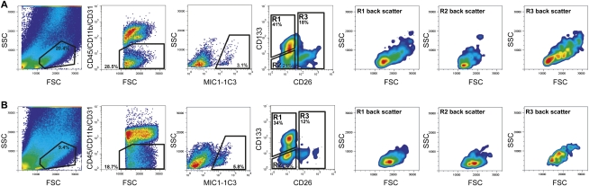
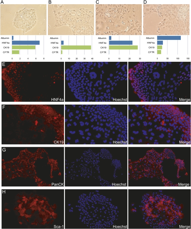
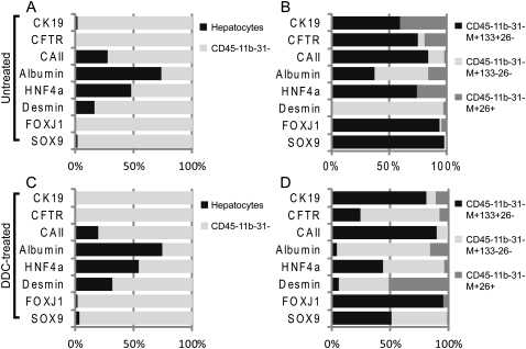
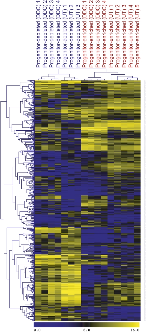
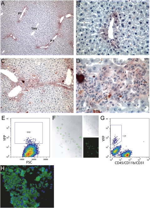
The molecular identification of adult hepatic stem/progenitor cells has been hampered by the lack of truly specific markers. To isolate putative adult liver progenitor cells, we used cell surface-marking antibodies, including MIC1-1C3, to isolate subpopulations of liver cells from normal adult mice or those undergoing an oval cell response and tested their capacity to form bilineage colonies in vitro. Robust clonogenic activity was found to be restricted to a subset of biliary duct cells antigenically defined as CD45(-)/CD11b(-)/CD31(-)/MIC1-1C3(+)/CD133(+)/CD26(-), at a frequency of one of 34 or one of 25 in normal or oval cell injury livers, respectively. Gene expression analyses revealed that Sox9 was expressed exclusively in this subpopulation of normal liver cells and was highly enriched relative to other cell fractions in injured livers. In vivo lineage tracing using Sox9creER(T2)-R26R(YFP) mice revealed that the cells that proliferate during progenitor-driven liver regeneration are progeny of Sox9-expressing precursors. A comprehensive array-based comparison of gene expression in progenitor-enriched and progenitor-depleted cells from both normal and DDC (3,5-diethoxycarbonyl-1,4-dihydrocollidine or diethyl1,4-dihydro-2,4,6-trimethyl-3,5-pyridinedicarboxylate)-treated livers revealed new potential regulators of liver progenitors.


Figures

Figure 1.
Figure 1.
FACS-based isolation of mouse liver NPC subsets. Cells isolated from normal (A) or DDC-injured (B) livers were enzymatically dispersed, antibody-labeled, and sorted by FACS. Successive gating showed sequential selection of cell-sized events (FSC vs. SSC), nonhematopoietic/endothelial events (CD45/CD11b/CD31 vs. FSC), and duct cells (MIC1-1C3 vs. FSC). Dead cells and debris were excluded using propidium iodide labeling and size exclusion, respectively. MIC1-1C3+ cells were divided into three subfractions (R1–R3) based on CD26 and CD133 expression; the size/granularity characteristics of the cells in R1–R3 are shown by backscattering on FSC/SSC.
Figure 2.
Figure 2.
Mouse liver cell colonies exhibit markers of both hepatocytic and ductal cell types. (A–D) Four representative individual colonies produced by FACS-sorted CD45/11b/31/MIC1-1C3+/26 cells are shown; the gene expression measurements for each colony are indicated below it. Expression data are presented as qRT–PCR-measured linearized ΔCt values relative to the mean of GAPDH and β-actin housekeeping genes. Albumin and HNF4a are markers of hepatocytic differentiation, whereas CK19 and CFTR are duct-associated. Phase contrast images are shown at 100×. (E–H) Additional colonies were grown on chamber slides and labeled with lineage-marking antibodies. Antibody labeling was visualized by Cy3 fluorescence and nuclei were labeled with Hoechst 33342. Magnification, 200×.
Figure 3.
Figure 3.
Mouse liver cell subset analysis reveals a duct subpopulation with progenitor gene expression. qRT–PCR results obtained from FACS-isolated hepatocyte and NPC populations were calculated as ΔCt values relative to the mean of GAPDH and β-actin; the relative expression in each cell subpopulation is indicated. The proportion of the total signal detected for a given gene in the populations being compared is indicated on the Y axis. (A,C) The expression levels of duct-, hepatocyte-, and progenitor-associated genes in hepatocytes and NPCs from normal (A) or DDC-injured (C) livers are compared. (B,D) The expression levels of duct-, hepatocyte-, and progenitor-associated genes in three subpopulations of MIC1-1C3+ NPCs from normal (B) or DDC-injured (D) livers are compared. Sox9 and FoxJ1 expression are restricted to the MIC1-1C3+/CD133+/CD26 duct subpopulation. Data from two replicate qRT–PCR analyses on material obtained from three separate cell isolation experiments were used.
Figure 4.
Figure 4.
Hierarchical clustering of microarray-assessed gene expression in mouse NPC subpopulations. Analysis of global gene expression profiles was performed using the 726 probes that were differentially expressed with a fold change of at least three between any cell type pair. (UT) Untreated; (DDC) oval cell induction.
Figure 5.
Figure 5.
Cells emerging from a DDC-induced ductular reaction are the progeny of a Sox9-expressing precursor. YFP expression was visualized in Sox9creERT2; R26RYFP mice 9 wk after tamoxifen injection. (A,B) Liver sections from control mice that received a normal diet during this interval. (C,D) Liver sections from mice that were DDC-treated to induce oval cell activation for the final 2 wk before harvest. (PT) Portal triad. Flow cytometric detection of YFP+ hepatocytes (E) and duct cells (G) recovered from the perfused liver of a tamoxifen-treated Sox9creERT2; R26RYFP mouse are shown. (F) Phase contrast and fluorescent imaging of sorted hepatocytes after overnight culture to confirm their identity and YFP fluorescence. (H) YFP+ colonies were initiated by clonogenic progenitors from these animals, indicating that the populations defined by Sox9 expression or surface marker labeling have the same identity. Magnifications: A,C, 100×; F,H,I, 400×; B,D, 500×.

References

    1. Barker N, Huch M, Kujala P, van de Wetering M, Snippert HJ, van Es JH, Sato T, Stange DE, Begthel H, van den Born M, et al. 2010. Lgr5(+ve) stem cells drive self-renewal in the stomach and build long-lived gastric units in vitro. Cell Stem Cell 6: 25–36 - PubMed
    1. Clayton E, Forbes SJ 2009. The isolation and in vitro expansion of hepatic Sca-1 progenitor cells. Biochem Biophys Res Commun 381: 549–553 - PubMed
    1. Dennis G Jr, Sherman BT, Hosack DA, Yang J, Gao W, Lane HC, Lempicki RA 2003. DAVID: database for annotation, visualization, and integrated discovery. Genome Biol 4: 3 doi: 10.1186/gb-2003-4-5-p3 - PubMed
    1. Dorrell C, Erker L, Lanxon-Cookson KM, Abraham SL, Victoroff T, Ro S, Canaday PS, Streeter PR, Grompe M 2008. Surface markers for the murine oval cell response. Hepatology 48: 1282–1291 - PMC - PubMed
    1. Duncan AW, Dorrell C, Grompe M 2009. Stem cells and liver regeneration. Gastroenterology 137: 466–481 - PMC - PubMed

Publication types