

Editorial
doi: 10.3324/haematol.2011.044628.
Ring sideroblasts and sideroblastic anemias
- PMID: 21632840
- PMCID: PMC3105636
- DOI: 10.3324/haematol.2011.044628
Item in Clipboard
Editorial
Ring sideroblasts and sideroblastic anemias
Haematologica.
2011 Jun.
No abstract available
Figures
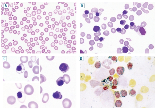
Representative peripheral blood and bone marrow smears from a patient with X-linked sideroblastic anemia. (A) Peripheral blood smear showing many hypochromic and microcytic cells. May-Grünwald-Giemsa (MGG), x1,000. (B) Bone marrow smear showing erythroid hyper plasia: erythroblasts are small with abnormal condensation of nuclear chromatin and ragged cytoplasm with ill-defined edges. MGG, x1,000. (C) Bone marrow smear showing erythroblasts with defective hemoglobinization (left) and erythroblasts containing multiple Pappenheimer bodies (right). MGG, x1,250. (D) Bone marrow smear. Perls’ stain shows that most erythroid precursors are ring sideroblasts with at least five positive granules disposed in a ring surrounding a third or more of the circumference of the nucleus. x1,250.
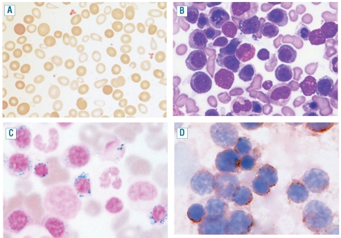
Representative peripheral blood and bone marrow smears from a patient with refractory anemia with ring sideroblasts. (A). Peripheral blood smear showing dimorphic red cells with a population of macrocytes and a population of hypochromic microcytes. MGG, x1,000. (B) Bone marrow smear showing a marked erythroid hyperplasia with megaloblastoid features. The rare granulocytic cells look normal. Upper right, a late erythroblast with defective hemoglobinization; lower right, an early erythroblast with vacuolated cytoplasm and a late erythroblast with Pappenheimer bodies. MGG, x1,000. (C) Bone marrow smear stained by Perls’ reaction showing several ring sideroblasts. MGG x1,250. (D) Bone marrow smear. Mitochondrial ferritin is detected in granules surrounding the nucleus. Immunoalkaline phosphatase reaction, MGG x1250.
Comment on
-
Missense SLC25A38 variations play an important role in autosomal recessive inherited sideroblastic anemia.Haematologica. 2011 Jun;96(6):808-13. doi: 10.3324/haematol.2010.039164. Epub 2011 Mar 10. Haematologica. 2011. PMID: 21393332 Free PMC article.
References
-
- Cazzola M, Invernizzi R. Sideroblastic anemias. In: Young NS, Gerson SL, High KA, editors. Clinical Hematology. Philadelphia: Mosby Elsevier; 2005. pp. 721–32.
-
- Mufti GJ, Bennett JM, Goasguen J, Bain BJ, Baumann I, Brunning R, et al. Diagnosis and classification of myelodysplastic syndrome: International Working Group on Morphology of myelodysplastic syndrome (IWGM-MDS) consensus proposals for the definition and enumeration of myeloblasts and ring sideroblasts. Haematologica. 2008;93(11):1712–7. - PubMed
-
- Cazzola M, Invernizzi R, Bergamaschi G, Levi S, Corsi B, Travaglino E, et al. Mitochondrial ferritin expression in erythroid cells from patients with sideroblastic anemia. Blood. 2003;101(5):1996–2000. - PubMed
-
- Cotter PD, May A, Li L, Al-Sabah AI, Fitzsimons EJ, Cazzola M, et al. Four new mutations in the erythroid-specific 5-aminolevulinate synthase (ALAS2) gene causing X-linked sideroblastic anemia: increased pyridoxine responsiveness after removal of iron overload by phlebotomy and coinheritance of hereditary hemochromatosis. Blood. 1999;93(5):1757–69. - PubMed
-
- Cazzola M, May A, Bergamaschi G, Cerani P, Ferrillo S, Bishop DF. Absent phenotypic expression of X-linked sideroblastic anemia in one of 2 brothers with a novel ALAS2 mutation. Blood. 2002;100(12):4236–8. - PubMed
Publication types
MeSH terms
LinkOut - more resources
Full Text Sources
Other Literature Sources
