. 2011 Oct 1;92(1):85-94.
doi: 10.1093/cvr/cvr159. Epub 2011 Jun 1.

Cardiac-specific mindin overexpression attenuates cardiac hypertrophy via blocking AKT/GSK3β and TGF-β1-Smad signalling

Affiliations

Cardiac-specific mindin overexpression attenuates cardiac hypertrophy via blocking AKT/GSK3β and TGF-β1-Smad signalling

Ling Yan et al. Cardiovasc Res. .

Abstract

Aims: Mindin is a secreted extracellular matrix protein, an integrin ligand, and an angiogenesis inhibitor, other examples of which are all key players in the progression of cardiac hypertrophy. However, its function during cardiac hypertrophy remains unclear. This study was aimed to identify the effect of mindin on cardiac hypertrophy and the underlying mechanisms.

Methods and results: A significant down-regulation of mindin expression was observed in human failing hearts. To further investigate the role of mindin in cardiac hypertrophy, we used cultured neonatal rat cardiomyocytes with gain and loss of mindin function and cardiac-specific Mindin-overexpressing transgenic (TG) mice. In cultured cardiomyocytes, mindin negatively regulated angiotensin II (Ang II)-mediated hypertrophic growth, as detected by [(3)H]-Leucine incorporation, cardiac myocyte area, and hypertrophic marker protein levels. Cardiac hypertrophy in vivo was produced by aortic banding (AB) or Ang II infusion in TG mice and their wild-type controls. The extent of cardiac hypertrophy was evaluated by echocardiography as well as by pathological and molecular analyses of heart samples. Mindin overexpression in the heart markedly attenuated cardiac hypertrophy, fibrosis, and left ventricular dysfunction in mice in response to AB or Ang II. Further analysis of the signalling events in vitro and in vivo indicated that these beneficial effects of mindin were associated with the interruption of AKT/glycogen synthase kinase 3β (GSK3β) and transforming growth factor (TGF)-β1-Smad signalling.

Conclusion: The present study demonstrates for the first time that mindin serves as a novel mediator that protects against cardiac hypertrophy and the transition to heart failure by blocking AKT/GSK3β and TGF-β1-Smad signalling.


Figures

Figure 1
Figure 1
Mindin expression is decreased in human failing hearts. (A) Representative western blots of mindin, ANP, and BNP in human failing hearts and donor hearts. (B) Real-time PCR analysis of mindin, ANP, and BNP in human failing hearts and donor hearts (n= 6). Values represent mean ± SEM. *P <0.01 compared with donor hearts.
Figure 2
Figure 2
Forced mindin expression attenuates the hypertrophic growth of cultured myocytes. (A) Representative western blots for ANP, BNP, and β-MHC after treatment with Ang II for the indicated times in neonatal rat cardiac myocytes infected with different adenoviruses. Reproducible results were obtained in three independent experiments. (B) Effects of mindin on [3H]-Leucine incorporation and cardiac myocyte area induced by Ang II treatment at the indicated time points. Cardiomyocytes were infected with Ad-GFP, Ad-mindin, Ad-shRNA, or Ad-shmindin for 24 h and then incubated with 1 µM Ang II for the indicated times. Values represent mean ± SEM. *P <0.01 compared with the Ad-GFP + Ang II group at time 0.
Figure 3
Figure 3
Generation of mice with cardiac-specific overexpression of mindin. (A) Representative blots for human mindin from various tissues of TG mice, as indicated. (B) Representative blots for transgenic mindin and endogenous mindin levels in heart tissue from five lines of both TG and WT mice.
Figure 4
Figure 4
Forced mindin expression in the heart produces resistance to cardiac remodelling in response to pressure overload or Ang II stimulation. (A and B) Statistical results for the HW/BW, HW/TL, and LW/BW ratios as well as myocyte cross-sectional areas (n= 100 cells per group) 4 weeks after AB surgery (A) or Ang II infusion (B) in WT and TG mice (n= 5–6 mice per group). (C and D) Protein expression levels of ANP, BNP, and β-MHC 4 weeks after AB surgery (C) or Ang II infusion (D) in WT and TG mice (n= 4). Left, representative blots; right, quantitative results. *P <0.01 for WT/sham or WT/saline values; P <0.01 for WT/AB or WT/Ang II after AB or Ang II infusion.
Figure 5
Figure 5
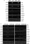
Mindin suppresses AKT/GSK3β/mTOR/FOXO signalling in vivo and in vitro. (A) Representative blots for AKT, GSK3β, mTOR, FOXO3A, and FOXO1 phosphorylation as well as their total protein expression levels 4 weeks following AB surgery or Ang II infusion in WT and TG (n= 4). (B) Representative blots for AKT, GSK3β, mTOR, FOXO3A, and FOXO1 phosphorylation and their total protein expression levels after treatment with Ang II for the indicated times in neonatal rat cardiomyocytes infected with Ad-GFP, Ad-mindin, Ad-shRNA, or Ad-shmindin. Reproducible results were obtained in three independent experiments.
Figure 6
Figure 6
Forced mindin expression attenuates fibrosis in response to pressure overload or Ang II stimulation. (A and B) Histological sections of the LVs of WT and TG mice (n= 6) were stained with PSR at 4 weeks after AB surgery (A) or Ang II infusion (B). Left, representative images; right, quantitative results. Scale bar: 32 µm. Fibrotic areas of the histological sections were quantified using an image analysis system. *P < 0.01 for WT/sham or WT/saline values; P < 0.01 for WT/AB or WT/Ang II after AB or Ang II infusion.

References

    1. Swynghedauw B, Delcayre C, Samuel JL, Mebazaa A, Cohen-Solal A. Molecular mechanisms in evolutionary cardiology failure. Ann N Y Acad Sci. 2010;1188:58–67. doi:10.1111/j.1749-6632.2009.05084.x. - DOI - PubMed
    1. Ashrafian H, Frenneaux MP, Opie LH. Metabolic mechanisms in heart failure. Circulation. 2007;116:434–448. doi:10.1161/CIRCULATIONAHA.107.702795. - DOI - PubMed
    1. Swynghedauw B. Molecular mechanisms of myocardial remodeling. Physiol Rev. 1999;79:215–262. - PubMed
    1. Jane-Lise S, Corda S, Chassagne C, Rappaport L. The extracellular matrix and the cytoskeleton in heart hypertrophy and failure. Heart Fail Rev. 2000;5:239–250. doi:10.1023/A:1009857403356. - DOI - PubMed
    1. Giancotti FG, Ruoslahti E. Integrin signaling. Science. 1999;285:1028–1032. doi:10.1126/science.285.5430.1028. - DOI - PubMed

Publication types

MeSH terms