. 2011 Jun 1;31(22):8046-58.
doi: 10.1523/JNEUROSCI.6671-10.2011.

Canonical Notch signaling is not necessary for prosensory induction in the mouse cochlea: insights from a conditional mutant of RBPjkappa

Affiliations

Canonical Notch signaling is not necessary for prosensory induction in the mouse cochlea: insights from a conditional mutant of RBPjkappa

Martín L Basch et al. J Neurosci. .

Abstract

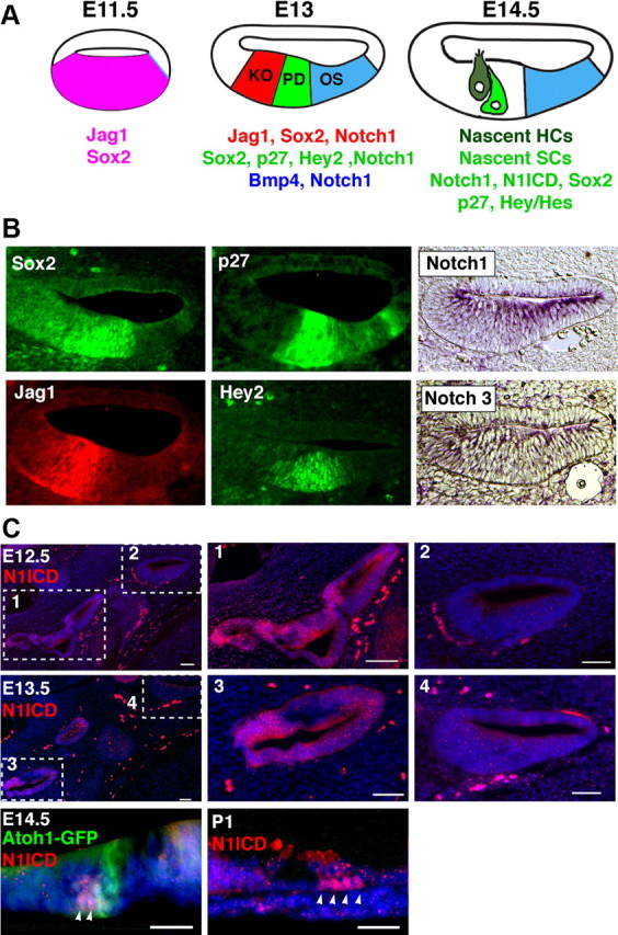
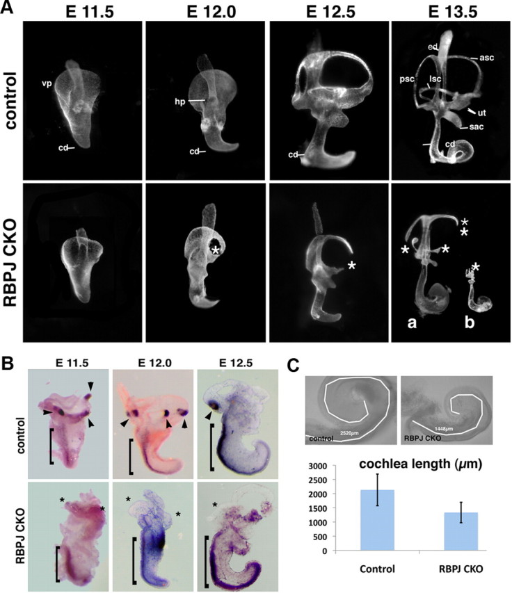
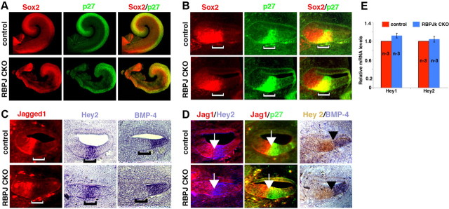
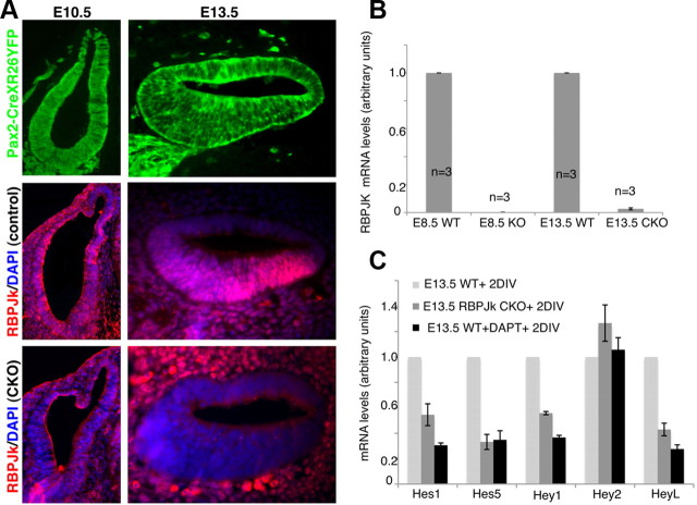
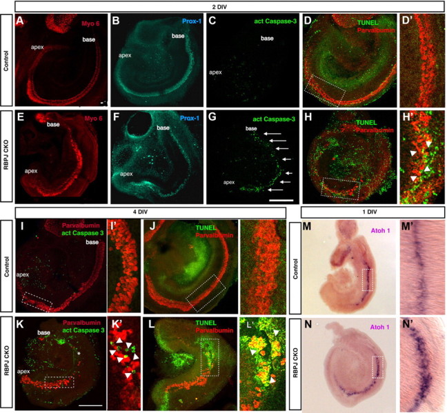
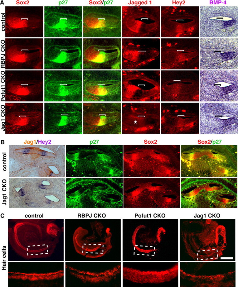
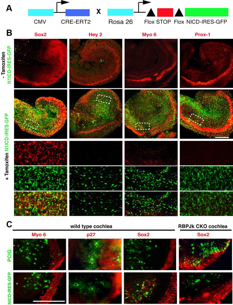
The mammalian organ of Corti consists of a highly organized array of hair cells and supporting cells that originate from a common population of prosensory progenitors. Proper differentiation of this complex cellular mosaic requires lateral inhibition mediated by Notch signaling. Several studies have implicated Notch signaling in the earlier induction of the prosensory domain that lies along the length of the cochlear duct, and which forms before the onset of hair cell and supporting cell differentiation. To investigate the role of Notch signaling in prosensory domain formation, we conditionally inactivated the transcriptional mediator of canonical Notch signaling, RBPjκ, throughout the inner ear. Although RBPjκ mutants have severe vestibular defects and a shortened cochlear duct, markers of the prosensory domain appear at the normal time and location in the cochlea of RBPjκ mutants. Despite the lack of RBPjκ, hair cell and supporting cell markers also appear at appropriate times in the cochlea, suggesting that RBPjκ is dispensable for differentiation of the cochlear sensory epithelium. However, we also observed that differentiating hair cells and supporting cells rapidly die in RBPjκ mutants, suggesting a requirement of RBPjκ for cell survival in this tissue. Finally, in contrast to the chick basilar papilla, ectopic activation of Notch signaling did not induce ectopic sensory patches in nonsensory regions of the cochlea. Our results indicate that canonical Notch signaling is not necessary for prosensory specification in the mouse cochlea, suggesting that other signaling pathways may specify this highly derived sensory organ.


Figures

Figure 1.
Figure 1.
Components of canonical Notch signaling are expressed in the developing cochlea. A, Schematic diagram illustrating the differentiation of the cochlea between E11.5 and E14.5. At E11.5, the ventral half of the cochlear duct expresses Jag1 and Sox2 (Ohyama et al., 2010). Between E12.5 and E13.0, this Jag1+;Sox2+ domain becomes restricted to Kölliker's organ (KO) on the neural side of the cochlear duct. A prosensory domain (PD) develops in the central portion of the duct, expressing Sox2, p27, and Hey2. It is bounded on the abneural side by the future outer sulcus (OS), which expresses Bmp4. At this stage, the entire cochlear duct expresses the Notch1 receptor. As hair cells (HC) and supporting cells (SC) differentiate after E13.5, activation of Notch1 can be observed in supporting cells (C). B, Sections through the cochlear duct of E13.5 embryos. p27kip1 and Sox2 immunostaining label the prosensory domain. Jagged1 and Hey2 label Kölliker's organ and the prosensory domain, respectively. In situ hybridization for Notch1 and Notch3 show expression throughout the ventral wall of the cochlea, with Notch3 being expressed at low levels. C, Notch1 receptor activation in the cochlea and vestibular system. At E12.5 and E13.5, the intracellular portion of the Notch1 receptor (N1ICD) can be detected in the vestibular system (boxes 1 and 3) but not in the cochlear duct on the same sections (boxes 2 and 4). Scale bars, 50 μm. At E14.5, N1ICD (arrowheads) can be detected in cells adjacent to differentiating Atoh1-expressing hair cells. These are likely to become supporting cells. By postnatal day 1, N1ICD is seen in a subset of supporting cells (arrowheads). Scale bar, 25 μm.
Figure 2.
Figure 2.
RBPjκ mutants develop severe inner ear morphological defects. A, Paint-filled inner ears from wild-type and RBPjκ CKO embryos between E11.5 and E13.5. The RBPjκ CKO displays no morphological defects at E11.5, but begins to show defects in semicircular canal formation at E12.0. At E13.5, two examples of mutant phenotype are shown, one (a) in which semicircular canal and vestibular sensory organ development are abnormal, and a second (b) in which the vestibular system is entirely absent. ed, Endolymphatic duct; asc, anterior semicircular canal; psc, posterior semicircular canal; lsc, lateral semicircular canal; ut, utricle; sac, saccule; cd, cochlear duct; vp, ventral plate; hp, horizontal plate; asterisks, canal truncations. B, Vestibular sensory patches are absent in RBPjκ mutants. Whole-mount in situ hybridization with a Bmp4 probe on inner ears dissected from E11.5–E12.5 wild-type and RBPjκ mutant embryos detects developing cristae in wild-type embryos (arrowheads) but not in RBPjκ mutants (asterisks). Bmp4 continues to be expressed along the abneural side of the cochlear duct in both wild-type and mutant ears (brackets). C, The RBPjκ CKO cochlea is shorter than wild-type. Bright field images of control and mutant E13.5 cochleas showing tracings of the cochlear duct using the length measurement function of Axiovision 4.7 software (white lines). The average cochlear length is displayed graphically (RBPjκ CKO, n = 7; control, n = 7; error bars show SD).
Figure 3.
Figure 3.
Normal patterning of the E13.5 cochlea in the absence of RBPjκ. A, Sox2 and p27kip1 antibody labeling of whole-mount cochleas from controls (top) or RBPjκ CKO (bottom) showing the prosensory domain. B, Sox2 and p27kip1 antibody labeling of sections of controls (top) or RBPjκ CKO (bottom) showing the prosensory domain (brackets). C, Jagged1 antibody staining and Hey2 and Bmp4 in situ hybridizations label Kölliker's organ, the prosensory domain, and outer sulcus, respectively. Top, Sections of controls; bottom, RBPjκ CKO; brackets, position of the prosensory domain. D, The border of the prosensory domain and Kölliker's organ (white arrows) and the border of the prosensory domain and the outer sulcus (black arrowheads) are maintained in controls (top) and RBPjκ CKO (bottom). Panels show Jag1 and p27kip1 antibody labeling, Bmp4 in situ hybridization, Hey2 in situ hybridization (left), and Hey2 immunostaining (right). E, mRNAs for the prosensory markers Hey1 and Hey2 are expressed at comparable levels in E13.5 wild-type and RBPjκ mutant cochleas as measured by qPCR.
Figure 4.
Figure 4.
Efficient conditional inactivation of RBPjκ by Pax2-Cre mice. A, Pax2-Cre transgenic mice were crossed to Rosa-YFP reporters to analyze the efficiency of recombination. Embryos were sectioned at E10.5 or E13.5 and all sections analyzed showed 100% recombination in the ventral otocyst and cochlear duct (top). The absence of RBPjκ protein was confirmed by immunostaining in controls and conditional mutants at the same ages (middle and bottom). B, RBPjκ mRNA levels of E13.5 conditional mutant cochleas or E8.5 null embryos were compared with controls by quantitative PCR. RBPjκ mRNA levels in the E13.5 conditional mutant cochleas are undetectable compared with wild-type. A similar result is seen in E8.5 RBPjκ-null embryos compared with wild-type embryos of the same age (all measurements were performed in triplicate; n = 3 in each case). C, E13.5 wild-type and RBPjκ mutant cochleas were cultured for 2 d and assayed for expression of Hes1, Hes5, Hey1, Hey2, and HeyL mRNA by qPCR. In addition, extra wild-type cultures were grown in the presence of DAPT for 2 d to inhibit Notch signaling. All Hes and Hey genes are significantly downregulated in RBPjκ mutants and DAPT-treated cultures, with the exception of Hey2. All measurements were performed in triplicate; N = 7 for Hes and Hes5; n = 4 for Hey1, Hey2, and HeyL. Error bars represent SEM.
Figure 5.
Figure 5.
Cochlear hair cells form and subsequently die in the absence of RBPjκ. A–L, E13.5 cochleas were cultured in a defined medium for 2 d (A–H) or 4 d (I–L). Cultures were stained with an antibody against activated caspase-3 (C, G, I, K) or TUNEL assays were performed (D, H, J, L) to analyze cell death. Hair cells were labeled with antibodies to Myo6 (A, E) or parvalbumin (D, H, I, K, J, L). Supporting cells were labeled with Prox1 antibodies (B, F). The sensory epithelium in RBPjκ mutants showed a significant increase in cell death (G, arrows; H′, K′, L′, arrowheads) compared with controls (C, D, D′, I, I′, J, J′). K, asterisk, Basal portion of the cochlea where hair cells died. D′, H′, I′, K′, J′, L′, Confocal planes through the dotted boxes in D, H, I, K, J, L, respectively, showing activated caspase-3 staining or positive TUNEL signal in the sensory epithelium of RBPjκ CKO cultures (H′, arrowheads). Scale bar, 200 μm. M, N, Atoh1 is expressed in the basal turn of the cochleas of E13.5 wild-type and RBPjκ mutants cultured for 24 h. The domain of Atoh1 is broader in RBPjκ mutants (N′) compared with wild-type cochleas (M′).
Figure 6.
Figure 6.
The prosensory domain of the cochlea is correctly specified in RBPJκ, Pofut1, and Jag1 mutants. A, Sections through the cochlear duct of E13.5 wild-type and RBPjκ, Pofut1, and Jag1 mutant embryos. The prosensory domain (brackets) was labeled with antibodies to p27Kip1 and in situ hybridization for Hey2, Kölliker's organ was labeled with antibodies to Jagged1, and the outer sulcus identified by in situ hybridization for Bmp4. Sox2 expression labeled both the prosensory domain and part of Kölliker's organ. All markers are expressed in normal patterns in all three mutants, with the exception of Jag1, which is absent in Jag1 CKO embryos (asterisk). B, Hey2 in situ hybridization in sections from control and Jagged1 CKO E14.5 embryos. Jagged1 immunostaining is shown in brown. Hey2 mRNA is still expressed in the differentiating prosensory domain (left). p27 and Sox2 immunostaining showing expression in the differentiating prosensory domain of control and Jagged1 CKO E14.5 embryos. C, E13.5 cochleas from control, RBPjκ, Pofut1, and Jag1 mutants were cultured for 2 d in vitro. Hair cell development was visualized by Myosin6 staining or an Atoh1-GFP reporter (Pofut1). Scale bar, 200 μm.
Figure 7.
Figure 7.
Activation of Notch signaling is not sufficient to induce prosensory or sensory fates in the cochlea. A, The diagram shows the tamoxifen-inducible system we used to express N1ICD throughout the cochlea. Upon addition of tamoxifen, Cre-ER removes a floxed stop cassette and allows expression of N1ICD and GFP driven by the Rosa26 promoter. B, E13.5 N1ICD-IRES-GFP, Cre-ER cochleas were cultured in the presence or absence of tamoxifen and allowed to develop for 48 h. We analyzed the expression of Sox2, Hey2, Myo6, and Prox1 by antibody staining (red). GFP expression was present in cells that expressed ectopic N1ICD. The bottom three rows show higher-magnification confocal planes of the dotted boxes above, showing the red channel (marker), green channel (GFP), and overlays. Only Sox2 was upregulated in response to ectopic N1ICD expression. C, E13.5 cochlear explants from wild-type mice were electroporated with a GFP control (PCIG) or an N1ICD-IRES-GFP construct, cultured, and examined for expression of Myosin 6 (96 h culture) or p27 and Sox2 (48 h culture). Although Sox2 is induced by N1ICD overexpression, no ectopic Myosin 6 or p27 cells are seen in the electroporated region. However, Sox2 was not induced when N1ICD was electroporated into RBPjκ mutant cochleas. Scale bars, 200 μm.

References

    1. Bok J, Chang W, Wu DK. Patterning and morphogenesis of the vertebrate inner ear. Int J Dev Biol. 2007;51:521–533. - PubMed
    1. Brooker R, Hozumi K, Lewis J. Notch ligands with contrasting functions: Jagged1 and Delta1 in the mouse inner ear. Development. 2006;133:1277–1286. - PubMed
    1. Cantos R, Cole LK, Acampora D, Simeone A, Wu DK. Patterning of the mammalian cochlea. Proc Natl Acad Sci U S A. 2000;97:11707–11713. - PMC - PubMed
    1. Chan SM, Weng AP, Tibshirani R, Aster JC, Utz PJ. Notch signals positively regulate activity of the mTOR pathway in T-cell acute lymphoblastic leukemia. Blood. 2007;110:278–286. - PMC - PubMed
    1. Chen P, Segil N. p27(Kip1) links cell proliferation to morphogenesis in the developing organ of Corti. Development. 1999;126:1581–1590. - PubMed

Publication types

MeSH terms

Substances

LinkOut - more resources