. 2011 Jun 1;31(22):8109-22.
doi: 10.1523/JNEUROSCI.1130-11.2011.

Girdin is an intrinsic regulator of neuroblast chain migration in the rostral migratory stream of the postnatal brain

Affiliations

Girdin is an intrinsic regulator of neuroblast chain migration in the rostral migratory stream of the postnatal brain

Yun Wang et al. J Neurosci. .

Abstract

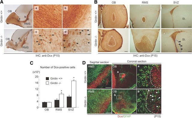
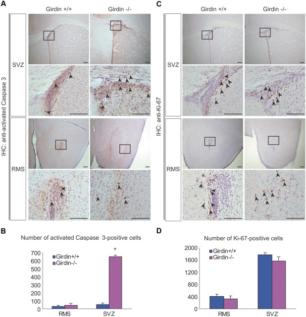
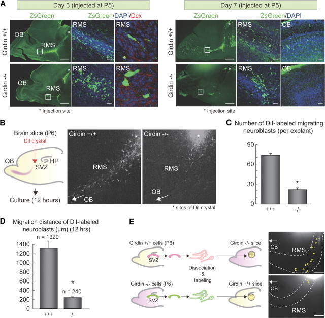
In postnatally developing and adult brains, interneurons of the olfactory bulb (OB) are continuously generated at the subventricular zone of the forebrain. The newborn neuroblasts migrate tangentially to the OB through a well defined pathway, the rostral migratory stream (RMS), where the neuroblasts undergo collective migration termed "chain migration." The cell-intrinsic regulatory mechanism of neuroblast chain migration, however, has not been uncovered. Here we show that mice lacking the actin-binding Akt substrate Girdin (a protein that interacts with Disrupted-In-Schizophrenia 1 to regulate neurogenesis in the dentate gyrus) have profound defects in neuroblast chain migration along the RMS. Analysis of two gene knock-in mice harboring Girdin mutants identified unique amino acid residues in Girdin's C-terminal domain that are responsible for the regulation of neuroblast chain migration but revealed no apparent requirement of Girdin phosphorylation by Akt. Electron microscopic analyses demonstrated the involvement of Girdin in neuroblast cell-cell interactions. These findings suggest that Girdin is an important intrinsic factor that specifically governs neuroblast chain migration along the RMS.


Figures

Figure 1.
Figure 1.
Defects in the development of the RMS and OB in Girdin−/− mice. A, Gross view of the brains of wild-type (left) and Girdin−/− (right) P15 mice, showing the decrease in OB size in Girdin−/− mice. B, C, Nissl-stained brain sections of P0 (B) and P15 (C) wild-type and Girdin−/− mice show significant deficits in the development of the RMS and the OB in Girdin−/− mice. Arrows indicate the width of the RMS. HP, Hippocampus. Scale bars, 1 mm. D, High-magnification views of the RMS (left) and the OB (right) of wild-type and Girdin−/− mice. GL, Glomerular layer; MC, mitral cell layer. E, Coronal sections through the OB (Ea), the RMS (Eb), and the SVZ (Ec) of wild-type (top) and Girdin−/− (bottom) P7 mice. The regions within the red boxes are shown at a higher magnification in adjacent panels. The far left panel shows schematic presentations of the cutting levels of coronal brain sections throughout this study. Scale bars, 200 μm.
Figure 2.
Figure 2.
Expression of Girdin in Dcx-positive migrating neuroblasts. A, Brain sections from wild-type (right) and Girdin−/− (far left) P15 mice were stained for Girdin. Brown staining denotes a positive Girdin signal. The regions within the black boxes (A and B) are shown at a higher magnification in adjacent panels. LV, Lateral ventricle; IHC, immunohistochemistry. Scale bars, 1 mm. B, X-gal staining of brain cryostat sections from wild-type (far left) and Girdin−/+ (right) P7 mice. The regions within the black boxes [the RMS and the hippocampus (HP)] are shown at a higher magnification in adjacent panels. Scale bars, 1 mm. C, Expression of Girdin in Dcx-positive type A neuroblasts but neither GFAP-positive astrocytes nor MASH1-positive transit amplifying cells in the SVZ. SVZ sagittal sections from wild-type and Girdin−/− P16 mice were double stained with the indicated antibodies for neuronal differentiation markers and Girdin antibodies. Enlarged images are shown in adjacent panels. D, Expression of Girdin in migrating neuroblasts in the RMS. RMS sagittal sections from wild-type P15 mice were double stained with anti-Girdin and indicated antibodies. Enlarged images are shown in insets. Scale bars, 50 μm.
Figure 3.
Figure 3.
Girdin deficiency impairs chain migration of RMS neuroblasts. A, P15 sagittal sections through the RMS were stained with anti-Dcx antibody, followed by DAB detection. The regions of the RMS within black boxes (a–d) are shown at a higher magnification on the right. In Girdin−/− mice, most of the Dcx-positive neuroblasts are migrating individually, and their leading processes are sometimes perpendicular to the direction of the stream (arrowheads). Scale bars: far left, 1 mm; a–d, 20 μm. B, P15 coronal sections through the OB, RMS, and SVZ were stained with anti-Dcx antibodies, showing dispersed RMS in Girdin−/− mice. RMS regions are indicated by dashed lines. Note that some neuroblasts were mismigrating toward the striatum in Girdin−/− mice (arrowheads). St., Striatum. Scale bars, 100 μm. C, Quantification of the distribution of Dcx-positive neuroblasts in wild-type and Girdin−/− P15 mice. The number of neuroblasts per high power field was counted and quantified. Five sections of the OB, RMS, and SVZ from three brains each of wild-type and Girdin−/− mice were evaluated. Data are expressed as the mean ± SEM, and comparison between wild-type and Girdin−/− mice was done by Student's t test. Asterisks indicate significant difference (p < 0.01) between wild-type and Girdin−/− mice. D, Gliosis of the RMS of Girdin−/− mice. Sagittal sections through the RMS (far left) and coronal sections through the OB (right) of wild-type and Girdin−/− P15 mice were stained with anti-Dcx (red) and anti-GFAP (green) antibodies. GFAP-positive cells (arrowheads) were frequently found in the RMS and intermixed with Dcx-positive neuroblasts in Girdin−/− mice. Scale bars, 100 μm. IHC, Immunohistochemistry.
Figure 4.
Figure 4.
Effects of Girdin deficiency on the apoptosis and the proliferation of SVZ neuroblasts. A, P15 coronal sections through the SVZ and RMS were stained with anti-activated Caspase-3 antibody, followed by DAB detection. Activated Caspase-3-positive cells (brown, highlighted by arrowheads) in the SVZ were numerous in Girdin−/− mice (right) compared with wild-type mice (left). B, Activated Caspase-3-positive cells were counted in six sections of the RMS or SVZ. Data are expressed as the mean ± SEM, and a comparison between wild-type and Girdin−/− mice was done by Student's t test. An asterisk indicates a significant difference (p < 0.01) between wild-type and Girdin−/− mice. C, P15 coronal sections through the SVZ and the RMS were stained with anti-Ki-67 antibody, followed by DAB detection. Ki-67-positive cells that progressed through the cell cycle (brown, highlighted by arrowheads) in these regions were similar between Girdin−/− mice (right) and wild-type mice (left). D, Ki-67-positive cells were counted in six sections of the RMS or SVZ. Data are expressed as the mean ± SEM, and a comparison between wild-type and Girdin−/− mice was done by Student's t test. No apparent significant difference between wild-type and Girdin−/− mice was found. IHC, Immunohistochemistry.
Figure 5.
Figure 5.
Girdin is essential for neuroblast chain migration in the RMS. A, Severe defects in the migration of newborn neuroblasts in Girdin−/− mice. A retrovirus bearing the ZsGreen cDNA was stereotactically injected into the SVZ of wild-type (top) and Girdin−/− (bottom) neonatal (P5) mice. Shown are sample confocal images of immunostaining with ZsGreen (green) and Dcx (red) antibodies. Chromosomal DNA was visualized with DAPI staining. RMS regions within white boxes are shown at a higher magnification in adjacent panels. At 3 and 7 d after the injection, the leading processes of the ZsGreen-positive neuroblasts are perpendicular to each other, leading to defects in neuroblast migration in Girdin−/− mice. In Girdin−/− mice, the majority of neuroblasts fail to reach the OB 7 d after retroviral injection (far right). Scale bars: far left, 1 mm; middle, 20 μm. B–D, Analysis of tangential neuroblast migration in brain slices. SVZ neuroblasts in acute brain slices isolated from wild-type and Girdin−/− P6 mice were labeled with DiI crystals placed in the SVZ (asterisks) and cultured for 12 h. The number of DiI-labeled migrating neuroblasts (C) and their migration distance (D) were quantified. In brain slices isolated from wild-type mice, the DiI-labeled neuroblasts tangentially migrated from the SVZ toward the OB in contrast to brain slices isolated from Girdin−/− mice, where the neuroblasts stayed within the SVZ and poorly migrated out from the SVZ. Asterisks indicate a significant difference (p < 0.0001) between wild-type and Girdin−/− mice. HP, Hippocampus. E, Cell-intrinsic function of Girdin in the migration of neuroblasts. Neuroblasts in the SVZ isolated from wild-type P6 mice were dissociated, labeled with DiI, and transplanted onto acute brain slices prepared from Girdin−/− mice (top). In a reciprocal experiment, SVZ neuroblasts from Girdin−/− mice were transplanted onto wild-type brain slices (bottom). Twenty-four hours after transplantation, fluorescent images show that Girdin−/− mice-derived neuroblasts failed to migrate toward the OB on the wild-type brain slices. Yellow arrowheads denote migrating neuroblasts.
Figure 6.
Figure 6.
Girdin is essential for neuroblast chain migration and chemoattraction. A, Expression of Girdin in Dcx-positive cultured neuroblasts. RMS explants from wild-type P6 mice were cultured in Matrigel for 24 h. The explants and emigrating neuroblasts were stained with anti-Girdin (green) and anti-Dcx (red) antibodies. Scale bars, 100 μm. B, RMS explants from wild-type (left) and Girdin−/− (right) P6 mice were cultured in Matrigel for 2 d. The explants and emigrating neuroblasts were stained with anti-GFAP (green) and anti-Dcx (red) antibodies. The transmitted differential interference contrast (DIC) images show that the neuroblasts migrate out from the explant as chains (yellow arrowheads) in wild-type mice, whereas cells in Girdin−/− mice failed to migrate out from the explants (red arrowheads) with only the extension of leading processes (white arrowheads). C, D, Time-lapse observation of neuroblasts emigrating from wild-type and Girdin−/− RMS explants cultured in Matrigel. The morphology of individual neuroblasts was observed in vitro by taking sequential DIC images of neuroblasts at 30 min intervals. Arrows and arrowheads denote cell bodies and leading processes of migrating neuroblasts, respectively. In neuroblasts from Girdin−/− RMS explants, the nuclei seem to stay in the proximity of the explant with only an extension of leading processes. Presented are representative images of two independent experiments. D, The velocity of neuroblasts migrating from Girdin+/+ and Girdin−/− RMS explants was quantified and analyzed. An asterisk indicates a significant difference (p < 0.0001) between wild-type and Girdin−/− mice. E, Girdin is essential for chemoattractive migration of cultured neuroblasts. RMS explants from wild-type (top) and Girdin−/− (bottom) P6 mice were cocultured with agarose beads (arrowheads) coated with the indicated growth factors for 2 d. The explants and emigrating neuroblasts were stained with anti-Dcx antibodies (magenta). F, G, Quantification of the effects of growth factors on neuroblast migration after Dcx immunostaining. F, Schematic illustration of the scoring principle: the number of cells within quadrant regions being proximal and distal against the beads were counted and quantified as shown in G (left). G, Right, Areas of migrating neuroblasts, which was defined as the sum of the pixels of the areas with positive Dcx staining, were evaluated and quantified. For each condition, a total of 10–15 explants were evaluated in three independent experiments. Data are expressed as the mean ± SEM, and comparisons between proximal and distal quadrants were analyzed by Student's t test. Asterisks indicate a significant difference (p < 0.01) between proximal and distal quadrants.
Figure 7.
Figure 7.
The BR region in the CT domain of Girdin, but not its phosphorylation by Akt, is responsible for regulation of neuroblast migration. A, Proposed domain structures of mouse Girdin. Girdin is composed of three domains: an N-terminal domain (NT) that binds to DISC1, a central coiled-coil domain, and a CT domain that binds to actin filaments and the Gα protein family (Enomoto et al., 2005; Ghosh et al., 2008; Jiang et al., 2008; Kitamura et al., 2008; Weng et al., 2010). An Akt phosphorylation site and a BR region are contained in the CT1 domain located in the first half of the CT domain. Shown in red characters are amino acid mutations introduced in the mutant mice generated in this study. B, Generation of SA and Basic-mut knock-in mutant mice. The mouse girdin gene consists of 33 coding exons, and exons 23–29 are shown here. Homologous recombination of the gene-targeting vector at the girdin locus (wild-type allele; top) was designed to insert a PGK-neo cassette into intron 25 and introduce indicated amino acid mutations that are encoded by exon 25. The structure of the targeted girdin allele (bottom) is shown. The position of the PCR-amplified genomic DNA probe used to screen ES cell colonies by Southern blotting is shown in the red box. Restriction enzyme sites for Southern blotting and predicted size of bands on Southern blotting are also shown. All data pertaining to mutant mice in this study were prepared in mutant mice without the neo gene. DTa, diphtheria toxin-A. C, Western blot analysis of brain lysates from wild-type and indicated mutant mice using anti-Girdin, anti-phospho-Girdin (P-Girdin), and anti-β-actin antibodies. WB, Western blot. D, Body weights and mortality rates in offspring from wild-type and Basic-mut mice. E, P15 sagittal brain sections through the OB, RMS, and hippocampus of SA and Basic-mut mice were Nissl stained (far left) and immunostained with Dcx antibodies (right). Subregions of the RMS and the DG within the black boxes (a–d) are shown at a high magnification in adjacent panels. In Basic-mut mice, but not SA mice, the migration of Dcx-positive neuroblasts was dysregulated both in the RMS and the DG, which seems to recapitulate the phenotype of Girdin−/− mice. IHC, Immunohistochemistry; HP, hippocampus. F, The BR region of Girdin is responsible for the regulation of migration in cultured neuroblasts. RMS explants from the SA and the Basic-mut P6 mice were cultured in Matrigel for 2 d. The explants and emigrating neuroblasts were stained with Dcx antibodies (magenta). Nuclei were visualized with DAPI staining (white).
Figure 8.
Figure 8.
Involvement of Girdin in neuroblast cell–cell interactions. A, Defects in cell–cell interactions of neuroblasts in Girdin−/− and Basic-mut mice. Selected coronal sections through the RMS from indicated mice were analyzed by electron microscopy. The regions in white boxes are shown at a higher magnification in adjacent panels. In wild-type mice (left), zonula adherens-like contact zones appear between closely apposed neuroblasts over a long distance (arrowheads), which seem to be absent or immature in Girdin−/− (middle) or Basic-mut (right, arrowheads) mice, respectively. N, Nuclei. B, C, Electron immunocytochemistry for the detection of Girdin localization in the RMS of wild-type (left), Girdin−/− (middle), and Basic-mut (right) mice. In wild-type mice, Girdin preferentially localizes to small zonula adherens-like contacts between neuroblasts (arrowheads). In contrast, in Basic-mut mice, Girdin seems to be localized to the cytoplasm apart from the plasma membrane (white arrowheads). C, Quantification of the percentage of membrane-localized immunoreactivity for Girdin in the neuroblasts of wild-type and Basic-mut mice. Girdin subcellular localization was classified into three different categories (both sides of cell–cell contacts; one side of cell–cell contacts; cytoplasm) based on the observation of Girdin immunoreactivity.
Figure 9.
Figure 9.
No apparent involvement of DISC1 and its interaction with Girdin in neuroblast migration in the RMS. A, Schematic diagram of the retroviral vector (pUEG) harboring control or DISC1 shRNAs used for in vitro birth-dating and genetic manipulation. LTR, Long terminal repeat. B–E, No apparent effects of DISC1 knockdown on the migration of newborn neuroblasts. Control or DISC1 shRNA-expressing retroviruses were injected into the SVZ of neonatal (P4) mice. Four (B, C) and seven (D, E) days after the retrovirus injection, the OB and RMS sagittal sections were prepared and immunostained (IS) with anti-GFP antibodies. Chromosomal DNA was visualized with DAPI staining. Shown in C and D is a quantification of the numbers of neuroblasts that reach the OB at the experimental time points. N.S., Not significant.
Figure 10.
Figure 10.
Cytoarchitectonic structure of the neocortex in Girdin−/− mice. A, Cortical layering and the distribution of Calbindin/Ctip2-positive interneuron in Girdin−/− mice. P15 sagittal sections of wild-type and Girdin−/− mice were stained with anti-Calbindin (left) and Ctip2 (right) antibodies, followed by DAB detection. On the right of each panel, the number of Calbinding/Ctip2-positive neurons in each layer was counted and quantified. An asterisk indicates a significant difference (p < 0.01) between wild-type and Girdin−/− mice. B, Distribution of Cajal-Retzius (CR) cells in Girdin−/− mice. E15 coronal sections of wild-type and Girdin−/− mice were stained with anti-Reelin antibody (left), and the number of Reelin-positive cells in the marginal zone in each hemisphere was counted and quantified (right), showing that Girdin−/− mice show a comparable distribution/positioning of CR cells as observed in wild-type mice. Arrowheads indicate Reelin expression in CR cells. C, Distribution of Reelin-positive neurons in Girdin−/− postnatal mice. P15 sagittal sections were stained with anti-Reelin antibody. Right, The cortex area of each section was roughly divided into three layers, and the number of Reelin-positive neurons in each layer was counted and quantified, showing no evident deficits in the distribution of Reelin-positive neurons in Girdin−/− mice. D, Defects in the development of cortical interneurons in Girdin−/− mice. P15 sagittal sections were stained with anti-GABA (far left), anti-parvalbumin (middle), and anti-Calretinin (far right) antibodies. On the right of each panel, the number of immunoreactive neurons was counted and quantified, showing impaired migration and/or differentiation of GABAergic cortical interneurons in Girdin−/− mice. Asterisks indicate a significant difference (p < 0.001) between wild-type and Girdin−/− mice. N.S., Not significant.

References

    1. Alcántara S, Ruiz M, D'Arcangelo G, Ezan F, de Lecea L, Curran T, Sotelo C, Soriano E. Regional and cellular patterns of reelin mRNA expression in the forebrain of the developing and adult mouse. J Neurosci. 1998;18:7779–7799. - PMC - PubMed
    1. Alvarez-Buylla A, García-Verdugo JM. Neurogenesis in adult subventricular zone. J Neurosci. 2002;22:629–634. - PMC - PubMed
    1. Anton ES, Ghashghaei HT, Weber JL, McCann C, Fischer TM, Cheung IM, Gassmann M, Messing A, Klein R, Schwab MH, Lloyd KC, Lai C. Receptor tyrosine kinase ErbB4 modulates neuroblast migration and placement in the adult forebrain. Nat Neurosci. 2004;7:1319–1328. - PubMed
    1. Belvindrah R, Hankel S, Walker J, Patton BL, Muller U. β1 integrins control the formation of cell chains in the adult rostral migratory stream. J Neurosci. 2007;27:2704–2717. - PMC - PubMed
    1. Chazal G, Durbec P, Jankovski A, Rougon G, Cremer H. Consequences of neural cell adhesion molecule deficiency on cell migration in the rostral migratory stream of the mouse. J Neurosci. 2000;20:1446–1457. - PMC - PubMed

Publication types

MeSH terms

LinkOut - more resources