. 2011 Jul 15;22(14):2509-19.
doi: 10.1091/mbc.E11-02-0157. Epub 2011 Jun 1.

A mechanism of Rap1-induced stabilization of endothelial cell--cell junctions

Affiliations

A mechanism of Rap1-induced stabilization of endothelial cell--cell junctions

Jian J Liu et al. Mol Biol Cell. .

Abstract

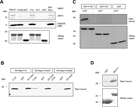
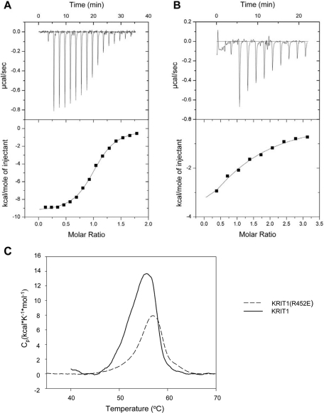
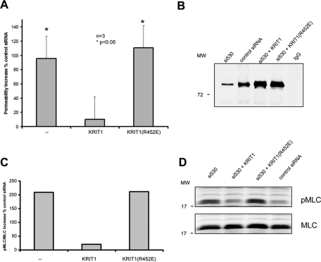
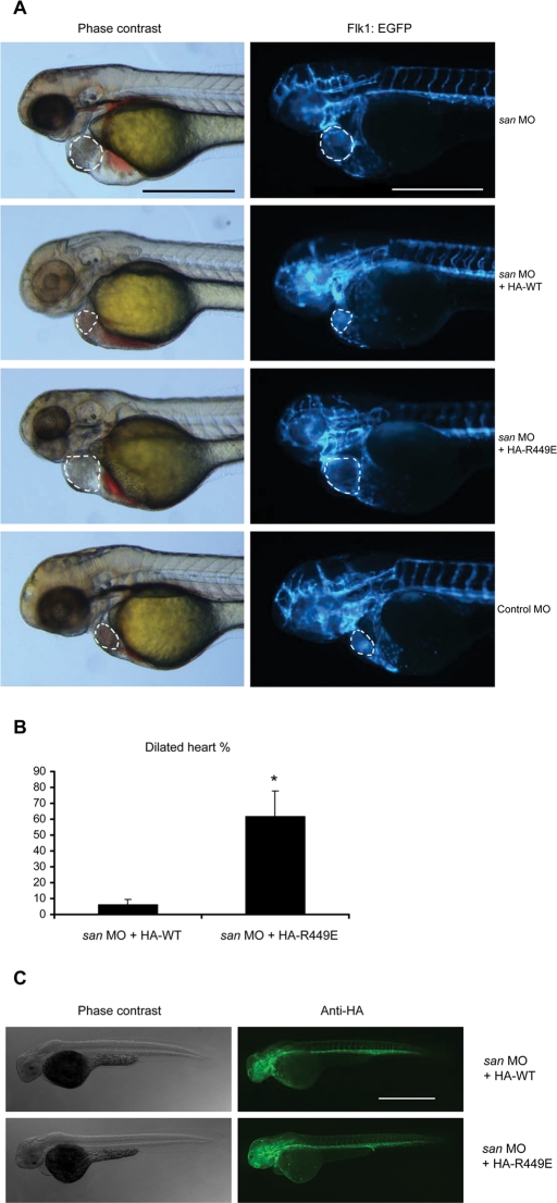
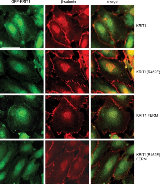
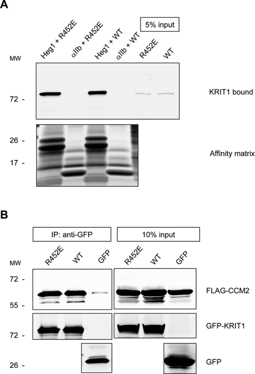
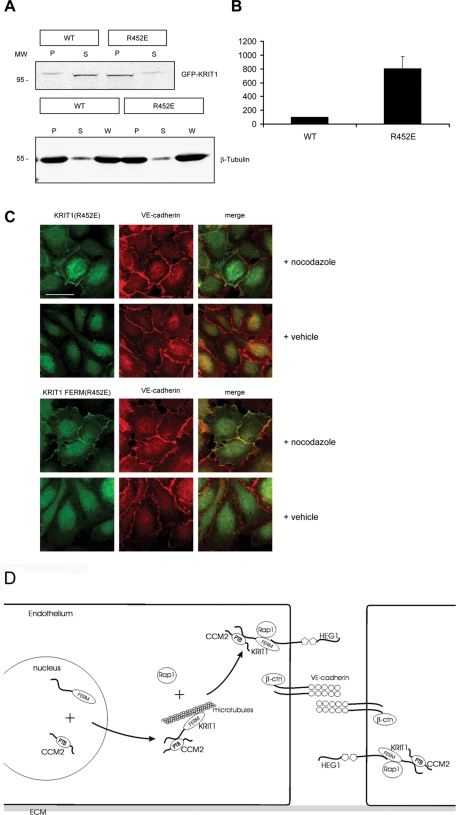
Activation of Rap1 small GTPases stabilizes cell--cell junctions, and this activity requires Krev Interaction Trapped gene 1 (KRIT1). Loss of KRIT1 disrupts cardiovascular development and causes autosomal dominant familial cerebral cavernous malformations. Here we report that native KRIT1 protein binds the effector loop of Rap1A but not H-Ras in a GTP-dependent manner, establishing that it is an authentic Rap1-specific effector. By modeling the KRIT1-Rap1 interface we designed a well-folded KRIT1 mutant that exhibited a ~40-fold-reduced affinity for Rap1A and maintained other KRIT1-binding functions. Direct binding of KRIT1 to Rap1 stabilized endothelial cell-cell junctions in vitro and was required for cardiovascular development in vivo. Mechanistically, Rap1 binding released KRIT1 from microtubules, enabling it to locate to cell--cell junctions, where it suppressed Rho kinase signaling and stabilized the junctions. These studies establish that the direct physical interaction of Rap1 with KRIT1 enables the translocation of microtubule-sequestered KRIT1 to junctions, thereby supporting junctional integrity and cardiovascular development.


Figures

FIGURE 1:
FIGURE 1:
KRIT1 is a Rap1-specific effector protein, and its interaction with Rap1 requires the F1 subregion of the FERM domain and the switch I domain of Rap1. (A) Recombinant bead-bound GST-Rap1A, GST-H-Ras, and GST were used to isolate recombinant KRIT1 and RalGDS from cell lysates. KRIT1 binds to Rap1A but not H-Ras or GST, whereas RalGDS binds to both Rap and Ras proteins (left). KRIT1 preferentially binds active Rap1A (GST-Rap1V12) and does not bind to an inactive Rap1 variant (GST-Rap1N17) or to GST only (right). GST blot is shown as loading control. Blots are representative; n = 3. Blots were cropped, and intervening lanes were removed using Adobe Photoshop. (B) KRIT1–Rap1A interaction involves residues within the switch 1 domain of Rap1A. Recombinant Rap1A (V12) from HEK293 cell lysate binds to GST-F123 (left). Single-alanine mutation on a switch 1 domain residue D38 (Rap1V12A38) does not bind to GST-F123 (right). Another switch 1 domain mutant (Rap1V12A37) dramatically reduces the binding to GST-F123 (middle). Blots are representative of three experiments. (C) KRIT1 binding to Rap1A is GTP dependent. Recombinant Rap1A from cell lysate binds to the KRIT1 FERM domain (GST-F123) only in the presence of GTP (10 mM). Binding of Rap1A to GST-KRIT1 FERM is lost on deletion of the F1 region, as binding to truncated FERM domain protein (GST-F23) is not observed (top). Expression of GST constructs is shown at bottom; blots are representative of three experiments. (D) F1 subregion of KRIT1 FERM domain is sufficient for Rap1A binding. Top, recombinant Rap1A (V12) binds to GST-F1 (right) but not GST alone (left). Bottom, equal loading of both GST fusion proteins as judged by SDS–PAGE and Coomassie Blue staining. Blots are representative of three experiments.
FIGURE 2:
FIGURE 2:
KRIT1 FERM domain homologous modeling reveals R452 residue mediating KRIT1–Rap1 interaction. (A) Homology model of KRIT1 FERM domain. Residues 410–736 from KRIT1 were modeled using the moesin crystal structure as the template. The modeled structure is formed by three subdomains that correspond to the F1 (orange), F2, and F3 (both in green) subregions of the FERM fold. F1 subregion structurally resembles the c-Raf RBD in a complex with Rap1. An arginine residue (R-452), whose corresponding residue in c-Raf (R-89) mediates the c-Raf–Rap1 binding, is also conserved in the KRIT1 F1 subregion. (B) A closer look at the modeled binding interface between KRIT1 F1 subregion (orange) and Rap1 (blue). The R452 residue in KRIT1 and switch 1 domain residues in Rap1 are shown in black. (C) The R452 residue is crucial for KRIT1–Rap1 binding. GST-F123 WT pulls down recombinant Rap1V12 from HEK293 cell lysate (second lane). The GST-F123(R452E) mutant form, which changes a basic residue to an acidic residue, does not bind to Rap1V12 (left lane). Bottom, the equal loading of both GST fusion proteins as judged by SDS–PAGE and Coomassie Blue staining. Blots are representative of five experiments.
FIGURE 3:
FIGURE 3:
Calorimetric characterization of KRIT1 FERM-domain binding to Rap1B bound to GMP-PNP, a GTP analogue. (A) Calorimetric titration of 400 μM Rap1B, out of the syringe, into 45 μM wild-type KRIT1 FERM domain in the sample cell. (B) Titration of 1.2 mM Rap1B into 45 μM KRIT1(R452E) FERM domain mutant protein. (C) KRIT1(R452E) FERM mutant does not disrupt protein folding. Differential scanning calorimetry results of wild-type and R452E FERM proteins both exhibit similar narrowly defined melting points, indicating that they are well folded.
FIGURE 4:
FIGURE 4:
KRIT1–Rap1 binding regulates HuVEC permeability. (A) Knockdown of KRIT1 expression by KRIT1 siRNA causes an approximately twofold increase in permeability (left). Reconstitution of recombinant WT KRIT1 reverses increased permeability (center). Recombinant KRIT1(R452E) mutant does not reverse increased permeability (right). Data shown are mean increased percentage over control siRNA in permeability ± SEM; n = 3. *p < 0,05 compared with control siRNA. (B) A representative blot of KRIT1 depletion and reexpression of recombinant proteins. More than 80% knockdown of KRIT1 is observed by immunoprecipitation and blotting. Reconstituted wild-type and RE (arginine 452 to glutamic acid) mutant KRIT1 proteins are expressed at equivalent levels. (C, D) Knockdown of KRIT1 expression causes an approximately threefold increase in pMLC/total MCL ratio (left). Reconstitution of recombinant WT KRIT1 reverses MLC phosphorylation increase (center). Recombinant KRIT1(R452E) mutant does not reverse increased pMLC level (right). Data shown are mean increased percentage over control siRNA; n = 2. (D) Immunoblots of pMLC (top) and total MLC (bottom) in HuVECs.
FIGURE 5:
FIGURE 5:
KRIT1–Rap1 interaction is required for normal zebrafish cardiovascular development in vivo. (A) Flk1:EGFP transgenic krit1-morphant (san MO) embryo showed enlarged heart phenotype. Coinjecting the cRNA encoding HA-tagged wild-type (WT) zebrafish krit1 protein reduced the heart size of injected fish, whereas fish coinjected with cRNA encoding HA-tagged Krit1(R449E) (RE, homologous to human R452E mutant) still exhibited a dilated heart phenotype. Dilated heart phenotype was scored by cardiac dilation in living fish in combination with a slower heart rate. Authentic dilation was verified by identifying the endocardium in the fluorescence images, which is indicated by the dotted lines. All microscopic images were taken at 48 hpf. Scale bars, 500 μm. (B) Bar graphs showing effects of Krit1 RE mutant on zebrafish cardiovascular development. Data are expressed as number of embryos with dilated heart phenotype divided by total number of embryos used per experiment times 100%, mean ± SD. Asterisk indicates p < 0.05 compared to san MO + HA-WT cRNA group. Data are from three independent experiments. Total number of animals used: 85 in san MO + HA-WT group, 120 in san MO + HA-RE group. (C) Lateral views of fixed animals at 48 hpf. Right, immunofluorescence images revealing that both HA-WT and HA-RE proteins were expressed at similar levels and throughout the entire fish. Scale bar, 500 μm.
FIGURE 6:
FIGURE 6:
Rap1 binding determines KRIT1 localization to endothelial cell–cell junctions. GFP-fused KRIT1 full-length protein (top) and FERM domain (third row from top) colocalize with β-catenin (red) in HuVEC cell–cell junctions. GFP-fused KRIT1(R452E) (second row from top) and KRIT1(R452E) FERM (bottom) mutants do not localize to HuVEC cell–cell junctions, whereas β-catenin (red) localizes to junctions. Confocal images are representative; n = 3. Bar, 50 μm.
FIGURE 7:
FIGURE 7:
Rap1 binding does not affect KRIT1 interactions with HEG1 or CCM2. (A) HEG1 cytoplasmic tail model protein binds to recombinant KRIT1 WT and R452E mutant proteins from HEK293 cell lysates. Integrin αIIb tail model protein does not bind to KRIT1. Bottom, the equal loading of tail proteins as judged by SDS–PAGE and Coomassie Blue staining. Blots are representative of three experiments. (B) Both GFP-fused KRIT1 WT and GFP-fused KRIT1(R452E) are associated with CCM2 at equivalent levels as assessed by coimmunoprecipitation and immunoblotting. Blots are representative of three experiments.
FIGURE 8:
FIGURE 8:
KRIT1–Rap1 interaction enables KRIT1 release from microtubules. (A) Top, KRIT1(R452E) (right two lanes) cosedimented with microtubules eightfold more than KRIT1 wild type (left two lanes). Bottom, immunoblotting of β-tubulin to show that the microtubule sedimentation is equivalent. P, pellet; S, supernatant; W, whole-cell lysate. (B) Quantification of KRIT1 cosedimented with microtubules. KRIT1 in the pellet was normalized to 100%. n = 3. (C) When microtubules were depolymerized by nocodazole, GFP-fused KRIT1(R452E) and KRIT1 FERM(R452E) localized to endothelial cell membranes and colocalized with junction marker VE-cadherin (red). Confocal images are representative; n = 3. Bar, 50 μm. (D) Mechanism of Rap1-mediated KRIT1 junctional localization. In endothelial cells, CCM2 binding enables KRIT1 egress from the nucleus. In the cytoplasm, KRIT1 is associated with microtubules. Rap1 binds KRIT1 and releases KRIT1 from microtubules. KRIT1 binds HEG1 to localize to endothelial cell–cell junctions, where it associates with junctional proteins, including β-catenin and VE-cadherin. Endothelial junction–localized KRIT1 controls junctional stability by suppressing RhoA activation. ECM, extracellular matrix.

Similar articles

Cited by

References

    1. Benkert P, Kunzli M, Schwede T. QMEAN server for protein model quality estimation. Nucleic Acids Res. 2009;37:W510–W514. - PMC - PubMed
    1. Beraud-Dufour S, Gautier R, Albiges-Rizo C, Chardin P, Faurobert E. Krit 1 interactions with microtubules and membranes are regulated by Rap1 and integrin cytoplasmic domain associated protein-1. FEBS J. 2007;274:5518–5532. - PMC - PubMed
    1. Bos JL. Linking Rap to cell adhesion. Curr Opin Cell Biol. 2005;17:123–128. - PubMed
    1. Cullere X, Shaw SK, Andersson L, Hirahashi J, Luscinskas FW, Mayadas TN. Regulation of vascular endothelial barrier function by Epac, a cAMP-activated exchange factor for Rap GTPase. Blood. 2005;105:1950–1955. - PubMed
    1. Emsley P, Lohkamp B, Scott WG, Cowtan K. Features and development of Coot. Acta Crystallogr D Biol Crystallogr. 2010;66:486–501. - PMC - PubMed

Publication types

MeSH terms