Comparative Study
. 2011 Sep;95(9):1323-30.
doi: 10.1136/bjo.2010.199216. Epub 2011 Jun 1.

C-reactive protein and complement factor H in aged human eyes and eyes with age-related macular degeneration

Affiliations
Comparative Study

C-reactive protein and complement factor H in aged human eyes and eyes with age-related macular degeneration

Imran A Bhutto et al. Br J Ophthalmol. 2011 Sep.

Abstract

Background: There is increasing evidence that inflammation and immune-mediated processes (complement activation) play an important role in age-related macular degeneration (AMD) pathogenesis. A genetic variation in the gene encoding complement factor H (CFH) and plasma levels of C-reactive protein (CRP), a systemic marker of subclinical inflammation, have consistently been shown to be associated with an increased risk for AMD. In the present study, we examined the immunolocalisation of CRP and CFH in aged control human donor eyes (n=10; mean age 79 years) and eyes with AMD (n=18; mean age 83 years).

Methods: Alkaline phosphatase immunohistochemistry was performed using polyclonal antibodies against CRP and CFH on cryopreserved tissue sections from disc/macular blocks. Three independent masked observers scored the reaction product (0-8).

Results: In aged control eyes, the retinal pigment epithelium/Bruch's membrane/choriocapillaris (RPE/BrM/CC) complex including intercapillary septa (ICS) had the most prominent immunostaining for CRP and CFH. CRP was significantly higher than controls in BrM/CC/ICS and choroidal stroma in early and wet AMD eyes (p<0.05). In contrast, CFH was significantly lower in BrM/CC/ICS complex of AMD choroids than in controls (p<0.05). Interestingly, CRP and CFH were significantly reduced in BrM/CC/ICS complex in atrophic area of macula in geographical atrophy (p<0.05). Drusen and basal laminar deposits were intensely positive for CRP and CFH.

Conclusion: These immunohistochemical findings show that changes in distribution and relative levels of CRP and CFH were evident in early and late AMD eyes. This suggests that high levels of CRP and insufficient CFH at the retina/choroid interface may lead to uncontrolled complement activation with associated cell and tissue damage. This study supports the hypothesis that inflammation and immune-mediated mechanisms are involved in the pathogenesis of AMD.


Figures

Figure 1
Figure 1
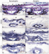
Retinal sections from an aged control, early, and wet AMD eyes are immunolabeled for CD34 (B,F,J), CRP (C,G,K) and CFH (D,H,L). The morphology of the retina is shown in the hematoxylin and eosin stained sections in A, E, and I. Endothelial cells of large retinal vessel (large arrow) and capillaries are intensely labeled for CD34 (B,F,J). Immunoreactivity for CRP (C) and CFH (D) is weak and associated with retinal vessel in aged control eye. Note that with severity of AMD, immunoreactivity for CRP (G,K) and CFH (H,L) is significantly increased. CFH and CRP in wet AMD are prominent within lumens (K,L) and in perivascular spaces around some large blood vessels (K). (scale bar=50μm in A-D; 30 μm in E-L)
Figure 2
Figure 2
Mean immunoreactivity scores ±SEM for CRP and CFH in retinal vessels of aged control (black), early AMD (white), and late AMD (gray) eyes. The scores for CRP were significantly higher in retinal vessels in wet AMD (arteries p≤0.0203; veins p=0.0026; and capillaries p=0.0009) (A) compared to aged controls and significantly lower in retinal artery (p=0.0026) and vein (p=0.0065) in atrophic and non-atrophic area in GA compared to aged controls (C). In contrast, there was no significant difference in scores for CFH in retinal vessels between wet AMD and aged controls (B) as well as between atrophic or non-atrophic areas in GA (D) and the aged controls. (*p<0.05 compared to control; unpaired Student’s t-Test analysis)
Figure 3
Figure 3
CRP and CFH immunoreactivity in choroid from aged control, early, and late wet AMD eyes. Periodic acid Schiff’s (PAS) and hematoxylin staining shows morphological features of the choroid from aged control (A), drusen (asterisk) in early AMD (B) and CNV (large arrow) anterior to RPE (open arrowhead) in wet AMD. The choriocapillaris (CC; small arrow) and large choroidal vessels are intensely labeled for CD34 and appear morphologically normal with broad lumens in aged control (D), whereas CC lumens appears irregular and constricted in early (E) and wet AMD (F). In aged control choroid, CRP (G) and CFH (J) are prominently localized to the CC, ICS and BrM (open arrowhead) and, to a lesser extent, in large choroidal vessels and stroma. CRP immunoreactivity is significantly increased in early (H) and late AMD (I) choroids compared to the aged control. CFH in early AMD (K) is comparable to aged control, whereas significantly decreased in wet AMD (L). Drusen are intensely positive for CRP and CFH (H and K). Note that in wet AMD the CNV (large arrow) area has more CRP and less CFH (I and L). Non-immune rabbit IgG yields a very weak to negative reaction product except in drusen (M, N, and O). (scale bar=20μm)
Figure 4
Figure 4
CRP and CFH immunoreactivity in AMD eye with GA. Note that the non-atrophic area (A) of choroid has RPE (arrowhead) and PAS-positive thickened BrM (double arrows) and no RPE in the atrophic area (B). CD34 localization demonstrates that the CC (arrow) is limited in the atrophic area compared to the non-atrophic area (C and D). In non-atrophic area, CRP is very intense in CC and blood vessels (asterisks) and diffuse throughout stroma (E). CFH is prominent in BrM, CC and ICS in nonatrophic areas (G). In the atrophic area, both antibodies yield reduced reaction product in BrM, CC and ICS (F and H). (scale bar=20μm)
Figure 5
Figure 5
Mean immunoreactivity scores ± SEM for CRP and CFH in choroidal structures of aged control (black), early AMD (white), and late AMD (gray) eyes. The scores for CRP were significantly higher in early AMD compared to controls: BrM p=0.0037, CC p<0.0001, ICS p<0.0001, arteries p=0.002, and veins p<0.001 (A). Scores were also significantly higher in wet AMD compared to controls: BrM p=0.05, CC p=0.027, and ICS p=0.024 (A). Scores were significantly lower for CRP in atrophic area in GA (C) compared to aged control and higher in non-atrophic regions compared to controls. In contrast, scores for CFH were significantly lower in choroidal structures in early and wet AMD (B). In early AMD, BrM (p=0.001), CC (p=0.014), and ICS (p<0.0001) had significantly less CFH than control subjects. In wet AMD, BrM, CC, ICS, arteries, veins, and stroma (p<0.0001, p<0.0001, p<0.0001, p<0.0001, p=0.002, p<0.001 respectively) had significantly less CFH than control subjects. Significant differences in CFH immunoreactivity were found in both non-atrophic (BrM, p<0.0001; CC, p=0.0013; and ICS, p<0.0001) and atrophic areas of GA (BrM, p=0.03; CC, p<0.0001; ICS, p<0.0001; artery, p=0.002) compared to the aged controls. (*p<0.05 by unpaired Student’s t-Test analysis between AMD and aged controls)

References

    1. Ambati J, Ambati BK, Yoo SH, et al. Age-related macular degeneration: etiology, pathogenesis, and therapeutic strategies. Surv Ophthalmol. 2003;48:257–293. - PubMed
    1. Johnson LV, Ozaki S, Staples MK, et al. A potential role for immune complex pathogenesis in drusen formation. Exp Eye Res. 2000;70:441–449. - PubMed
    1. Johnson LV, Leitner WP, Staples MK, et al. Complement activation and inflammatory processes in drusen formation and age related macular degeneration. Exp Eye Res. 2001;73:887–896. - PubMed
    1. Crabb JW, Miyagi M, Gu X, et al. Drusen proteome analysis: an approach to the etiology of age-related macular degeneration. Proc Natl Acad Sci USA. 2002;99:14682–14687. - PMC - PubMed
    1. Hageman GS, Mullins RF, Russell SR, et al. Vitronectin is a constituent of ocular drusen and the vitronectin gene is expressed in human retinal pigmented epithelial cells. FASEB J. 1999;13:477–484. - PubMed

Publication types

MeSH terms