Review
. 2011 Jun;121(6):2065-73.
doi: 10.1172/JCI45961. Epub 2011 Jun 1.

The three R's of lung health and disease: repair, remodeling, and regeneration

Affiliations
Review

The three R's of lung health and disease: repair, remodeling, and regeneration

Michael F Beers et al. J Clin Invest. 2011 Jun.

Abstract

All tissues and organs can be classified according to their ability to repair and regenerate during adult homeostasis and after injury. Some exhibit a high rate of constant cell turnover, while others, such as the lung, exhibit only low-level cell regeneration during normal adult homeostasis but have the ability to rapidly regenerate new cells after injury. Lung regeneration likely involves both activation of progenitor cells as well as cell replacement through proliferation of remaining undamaged cells. The pathways and factors that control this process and its role in disease are only now being explored. In this Review, we will discuss the connection between pathways required for lung development and how the lung responds to injury and disease, with a particular emphasis on recent studies describing the role for the epithelium in repair and regeneration.


Figures

Figure 1
Figure 1. Early patterning and morphogenesis of the anterior foregut and origin of epithelial cell lineages in the lung.
(A) Diagram of the early anterior foregut development, showing the relationship between Wnt2/2b and Fgf10, which are expressed in the early ventral anterior mesoderm surrounding the foregut. Wnt2/2b act upstream of Fgf10 (and other factors including Bmp4) to induce lung specification in the most ventral aspect of the anterior foregut endoderm, as noted by expression of Nkx2.1. (B) As lung development ensues, the endoderm goes through extensive and reiterated branching, setting up the proximal-distal patterning of the airways. This results in distinct progenitors located within the proximal (Sox2+) and distal (Sox9, Id2, Foxp2) airway epithelium. However, the distal Id2+ progenitors can generate proximal epithelium up to E13.5 of mouse development (B, arrows) (6).
Figure 2
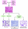
Figure 2. The 3 R’s of lung homeostasis.
Schematic model of injury-repair responses in the distal lung. In many cases of parenchymal lung diseases, including pulmonary fibrosis, emphysema, and pulmonary hypertension, both intrinsic (e.g., genetic mutations, mechanical stress, immunological signaling) and extrinsic (e.g., toxins, oxidants) sources of injury combine in a “multiple hit” process to produce resident cell (epithelial or endothelial) dysfunction. Local responses by affected cell populations can preserve normal homeostasis (repair), but, if these are unsuccessful, resident cell death ensues, and a decision either to regenerate (restoring normal architecture and physiology) or to remodel (producing the resultant dysfunctional disease state) is initiated. BPD, bronchopulmonary dysplasia.
Figure 3
Figure 3. The epithelial-mesenchymal wound model for lung injury and repair.
In the initiation stage, multiple microinjuries damage and activate alveolar epithelial cells. In the amplification stage, the resulting ER stress and other activating signals in AEC cells can promote release of chemokines/cytokines, promoting migration of inflammatory effector cells into the distal lung. Persistent activation and/or ongoing ER stress can result in apoptosis and AEC cell drop-out. In the response stage, in order to restore the denuded epithelial surface, repair via reepithelialization is attempted using either local proliferation/transdifferentiation of AT2 cells or regional expansion of progenitor cell populations such as BASCs (regeneration). In the absence of successful reepithelialization (repair or regeneration), remodeling is initiated. In a fibrotic phenotype (e.g., IPF), the local milieu (including high TGF-β and high VEGF levels) promotes proliferation of fibroblasts, activation of myofibroblasts, increases in basement membrane disruption, and neovascularization, with resulting formation of a collagen scar. In emphysema, under different local conditions (including low VEGF and low TGF-β levels), airspace enlargement takes place.
Figure 4
Figure 4. Cell lineages involved in repair and regeneration of the adult lung epithelium.
(A) In the adult proximal or bronchiolar epithelium, cells expressing Sox2, p63, and Krt5 are thought to be progenitors capable of regenerating injured or denuded airway epithelium. Whether these markers denote unique, distinct, or overlapping progenitor populations is unknown, but data show that p63 and Krt5 mark a more proximal population of cells than Sox2 alone, which is expressed throughout tracheal and bronchiolar regions (15, 70). Notch signaling is known to regulate the decision to generate secretory lineages (i.e., Scgb1a1+) versus neuroendocrine cells within the proximal airways. The secretory lineage has also been shown to generate both ciliated epithelium (β-tubulin IV+) as well as goblet cells (Muc5A/C+) after injury to the bronchiolar epithelium. (B) In the alveolar epithelium, less is known about the relationship between the two major cell types, AEC1 and AEC2 cells. There is some in vitro evidence that AEC2 cells can act as progenitors to repopulate lost AEC1 and AEC2 cells after injury (63). Additional genetic and molecular data are needed to assess whether there is a resident alveolar progenitor or whether AEC2 cells are facultative progenitors. Aqp5, aquaporin 5; CGRP, Calcitonin gene–related peptide; Pgp9.5, protein gene product 9.5; T1α, podoplanin.

References

    1. Cardoso WV, Lu J. Regulation of early lung morphogenesis: questions, facts and controversies. Development. 2006;133(9):1611–1624. doi: 10.1242/dev.02310. - DOI - PubMed
    1. Cardoso WV, Whitsett JA. Resident cellular components of the lung: developmental aspects. Proc Am Thorac Soc. 2008;5(7):767–771. doi: 10.1513/pats.200803-026HR. - DOI - PMC - PubMed
    1. Morrisey EE, Hogan BL. Preparing for the first breath: genetic and cellular mechanisms in lung development. Dev Cell. 2010;18(1):8–23. doi: 10.1016/j.devcel.2009.12.010. - DOI - PMC - PubMed
    1. Giangreco A, Reynolds SD, Stripp BR. Terminal bronchioles harbor a unique airway stem cell population that localizes to the bronchoalveolar duct junction. Am J Pathol. 2002;161(1):173–182. - PMC - PubMed
    1. Hong KU, Reynolds SD, Watkins S, Fuchs E, Stripp BR. Basal cells are a multipotent progenitor capable of renewing the bronchial epithelium. Am J Pathol. 2004;164(2):577–588. doi: 10.1016/S0002-9440(10)63147-1. - DOI - PMC - PubMed

Publication types

MeSH terms

Substances