

Standardization in digital pathology: Supplement 145 of the DICOM standards
- PMID: 21633489
- PMCID: PMC3097525
- DOI: 10.4103/2153-3539.80719
Standardization in digital pathology: Supplement 145 of the DICOM standards
Abstract
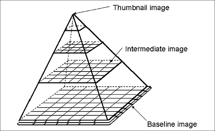
As digital slides need a lot of storage space, lack of a singular method to acquire and store these large, two-dimensional images has been a major stumbling block in the universal acceptance of this technology. The DICOMS Standard Committee Working Group 26 has put in a tremendous effort to standardize storage methods so that they are more in line with currently available PACS in most hospitals for storage of radiology images. A recent press release (Supplement 145) of these standards was hailed by one and all involved in the field of digital pathology as it will make it easier for hospitals to integrate digital pathology into their already established systems without adding too much overhead costs. Besides, it will enable different vendors developing the scanners to upgrade their products to storage systems that are common across all systems.
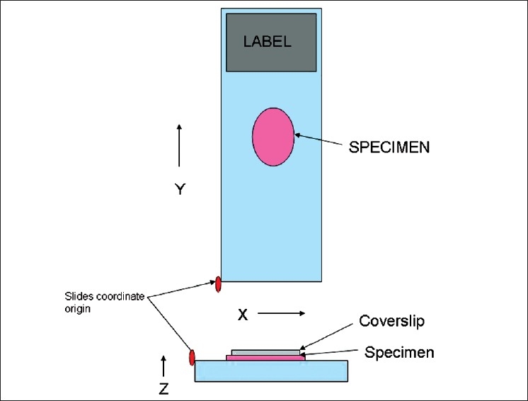
Keywords: DICOM; Digital pathology; z-planes.
Figures
References
-
- Supplement 145: Whole Slide Microscopic Image IOD and SOP Classes. Digital Imaging and Communications in Medicine (DICOM) 2010 Sep
-
- Le Bozec C, Henin D, Fabiani B, Schrader T, Garcia-Rojo M, Beckwith B. Refining DICOM for pathology--progress from the IHE and DICOM pathology working groups. Stud Health Technol Inform. 2007;129:434–8. - PubMed
-
- Daniel C, García Rojo M, Bourquard K, Henin D, Schrader T, Della Mea V, et al. Standards to support information systems integration in anatomic pathology. Arch Pathol Lab Med. 2009;133:1841–9. - PubMed
-
- Kahn CE, Jr, Carrino JA, Flynn MJ, Peck DJ, Horii SC. DICOM and radiology: Past, present, and future. J Am Coll Radiol. 2007;4:652–7. - PubMed
LinkOut - more resources
Full Text Sources
Research Materials
