. 2011;6(5):e20166.
doi: 10.1371/journal.pone.0020166. Epub 2011 May 25.

Neutralising antibodies against ricin toxin

Affiliations

Neutralising antibodies against ricin toxin

Julie Prigent et al. PLoS One. 2011.

Abstract

The Centers for Disease Control and Prevention have listed the potential bioweapon ricin as a Category B Agent. Ricin is a so-called A/B toxin produced by plants and is one of the deadliest molecules known. It is easy to prepare and no curative treatment is available. An immunotherapeutic approach could be of interest to attenuate or neutralise the effects of the toxin. We sought to characterise neutralising monoclonal antibodies against ricin and to develop an effective therapy. For this purpose, mouse monoclonal antibodies (mAbs) were produced against the two chains of ricin toxin (RTA and RTB). Seven mAbs were selected for their capacity to neutralise the cytotoxic effects of ricin in vitro. Three of these, two anti-RTB (RB34 and RB37) and one anti-RTA (RA36), when used in combination improved neutralising capacity in vitro with an IC(50) of 31 ng/ml. Passive administration of association of these three mixed mAbs (4.7 µg) protected mice from intranasal challenges with ricin (5 LD(50)). Among those three antibodies, anti-RTB antibodies protected mice more efficiently than the anti-RTA antibody. The combination of the three antibodies protected mice up to 7.5 hours after ricin challenge. The strong in vivo neutralising capacity of this three mAbs combination makes it potentially useful for immunotherapeutic purposes in the case of ricin poisoning or possibly for prevention.


Conflict of interest statement

Competing Interests: The authors have declared that no competing interests exist.

Figures

Figure 1
Figure 1. Immunoblot of ricin identified by several anti-ricin antibodies.
2 µg of ricin was migrated in 15% SDS-PAGE and blotted onto PVDF membrane. Primary monoclonal antibodies obtained against the A or B chain were incubated for 1 h to bind to ricin. A secondary antibody HRP-conjugated anti-mouse IgG (diluted 1/2000) was added and proteins were detected after 10 min by chemiluminescence (ECL) using a VersaDoc imaging system (Bio-Rad). Two lanes are shown for each antibody, corresponding to the migration of ricin and of molecular weight markers (two lines, 37 and 20 kDa), respectively.
Figure 2
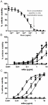
Figure 2. Ricin toxicity and antibody neutralising effect in vitro using a viability assay with Jurkat cells.
(A) Evaluation of ricin toxicity with Jurkat cells. Ricin (0–100 ng/ml) was incubated with 2×10⁴ cells/ml and cell viability was assessed by means of luminescence assay using the Cell titer Glo luminescence kit (Promega). (B) Neutralisation assay of ricin using anti-A chain antibodies (RA30: •; RA35: ▪; RA36: ▴; RA32: ▾ and RA33: ♦). (C) Neutralisation assay of ricin using anti-B chain antibodies (RB18: ◊; RB27: Δ; RB34: ○ and RB37: □). For Figures (B) and (C), 0.1 ng/ml ricin was pre-incubated with 0–10 µg/ml antibody and then exposed to 2×10⁴ cells/ml for 72 h before assessment of cell viability in the same way as in Figure (A).
Figure 3
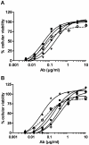
Figure 3. Combination neutralising effect of antibodies against ricin in vitro.
(A) Combination of pairs of antibodies, with RB34 as control (RB34: ○; RB34/RB37: ▴; RB34/RA36: ♦; RB37/RA36: ▾; RB27/RB37: ▪). (B) Combination of three or four antibodies, with RB34 as control (RB34: ○; RB27/RB37/RA36: •; RB34/RB37/RA36: ▴; RB34/RB37/RA36/RA32: +; RB34/RB37/RA36/RA33: ▪; RB34/RB37/RA36/RA35: ♦). Antibodies were premixed in equimolar ratio at several concentrations (0–10 µg/ml) and incubated with ricin (0.1 ng/ml) before exposure to Jurkat cells. Cell viability was assessed by means of luminescence assay using a Cell titer Glo luminescence kit (Promega).
Figure 4
Figure 4. Pharmacokinetic study of RB34, RB37 and RA36 in mice.
Purified antibody (50 µg) was injected intraperitoneally into Swiss mice (n = 4). Mice were sacrificed at different times to calculate plasma concentration of mAbs: RB34 (▪); RB37 (▴) and RA36 (•), using an immunoassay.
Figure 5
Figure 5. In vivo neutralising activity of anti-ricin antibodies combination pre-incubated with ricin (A) Survival curve.
CD1 mice were intranasally challenged with 5 LD50 of ricin alone (Δ) or pre-incubated with antibodies: several doses of RA36, RB34 and RB37 mixture (Abs) were assessed to obtain antibody/ricin molar ratios of 2 (▪), 5 (•), 10 (▴) and 20 (♦), compared with a nonspecific antibody (ns Ab) in the same concentration (R = 2 (+), R = 5 (X), R = 10 (○), R = 20 (□)). 50 µl of ricin-antibody complex was administered per mouse and mortality was monitored for 21 days. (B) Weight change. In the same experiment, mice were weighed at 0, 2, 7, 14 and 21 days. The percentage of the D0 weight (100%) was calculated, with female CD1 mice as control (▾). Mice injected with ricin alone (Δ) or with mixtures of specific antibodies at R = 2 (▪); R = 5 (•); R = 10 (▴) and R = 20 (♦). The data are representative of two independent experiments.
Figure 6
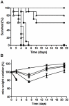
Figure 6. In vivo neutralising activity of anti-ricin antibodies combination administered after ricin challenge.
(A) Survival curve. CD1 mice were intranasally challenged with 5 LD50 of ricin alone (Δ) or ricin followed by intravenous injection of 5 mg/kg of antibodies 10 min (▪), 1 h (•), 5 h (♦), 7.5 h (▴), 10 h (○) and 24 h (□) after challenge. (B) Weight change. In the same experiment, mice were weighed at 0, 2, 7, 14 and 21 days, taking the weight at day zero as reference (100%), for female CD1 mice as a control (▾), mice injected with ricin (Δ), or ricin followed by intravenous injection of 5 mg/kg of antibodies 10 min (▪), 1 h (•), 5 h (♦), 7.5 h (▴), 10 h (○) and 24 h (□) after challenge. The data are representative of two independent experiments.
Figure 7
Figure 7. In vivo evaluation of each neutralising anti-ricin antibody administered after ricin challenge.
(A) Survival curve. CD1 mice were intranasally challenged with 5 LD50 of ricin alone (Δ) or ricin followed by intravenous injection of 5 mg/kg of RB34 antibody (▪), RB37 antibody (•), or RA36 antibody (□) 1 h after ricin challenge. (B) Weight change. In the same experiment, mice were weighed at 0, 2, 7, 14 and 21 days, taking the weight at day zero as reference (100%), for female CD1 mice as a control (▾), mice injected with ricin (Δ), or ricin followed by intravenous injection of 5 mg/kg of mouse weight of RB34 antibody (▪), RB37 antibody (•), or RA36 antibody (□) 1 hour after ricin challenge.
Figure 8
Figure 8. Competition of lactose with antibodies binding to ricin.
Neutralising anti-RTB antibodies RB34 (□) and RB37 (•) and a non neutralising antibody (RB18) used as control (▴) were incubated at 75 ng/ml with increasing concentrations of lactose (from 0 to 10 mM) and biotin-labeled RTB (25 ng/ml) in a microtiter plate coated with a polyclonal anti-mouse antibody. Antibodies binding to ricin were further revealed using streptavidin-AChE and Ellman's reagent. Absorbance was measured at 414 nm. B and B0 correspond to the absorbance obtained with or without competitor, respectively, allowing to calculate B/B0 (expressed as %) for each point.

References

    1. Audi J, Belson M, Patel M, Schier J, Osterloh J. Ricin poisoning: a comprehensive review. JAMA. 2005;294:2342–2351. - PubMed
    1. Bigalke H, Rummel A. Medical aspects of toxin weapons. Toxicology. 2005;214:210–220. - PubMed
    1. Binder P, Attre O, Boutin JP, Cavallo JD, Debord T, et al. Medical management of biological warfare and bioterrorism: place of the immunoprevention and the immunotherapy. Comp Immunol Microbiol Infect Dis. 2003;26:401–421. - PubMed
    1. Burnett JC, Henchal EA, Schmaljohn AL, Bavari S. The evolving field of biodefence: therapeutic developments and diagnostics. Nat Rev Drug Discov. 2005;4:281–297. - PMC - PubMed
    1. Cleveland DW, Fischer SG, Kirschner MW, Laemmli UK. Peptide mapping by limited proteolysis in sodium dodecyl sulfate and analysis by gel electrophoresis. J Biol Chem. 1977;252:1102–1106. - PubMed

Publication types

MeSH terms