. 2011;6(5):e20173.
doi: 10.1371/journal.pone.0020173. Epub 2011 May 25.

A role for hemopexin in oligodendrocyte differentiation and myelin formation

Affiliations

A role for hemopexin in oligodendrocyte differentiation and myelin formation

Noemi Morello et al. PLoS One. 2011.

Abstract

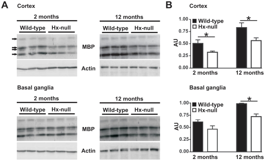
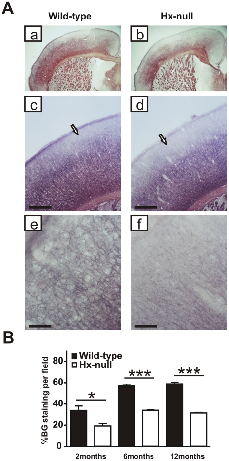
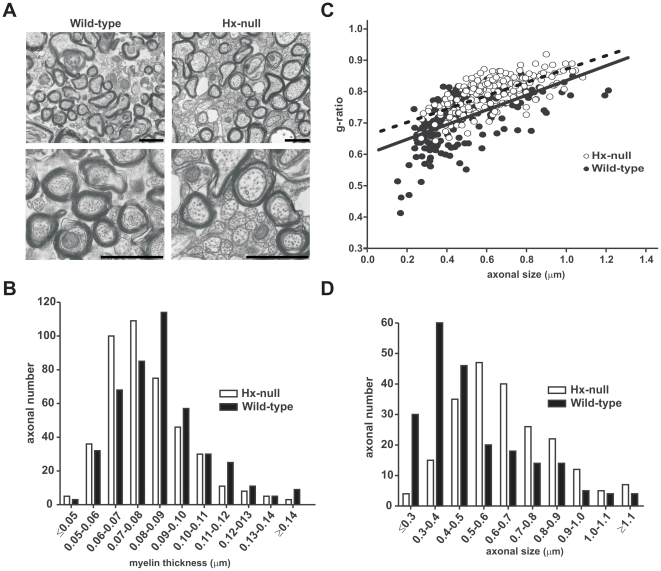
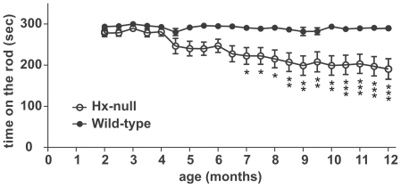
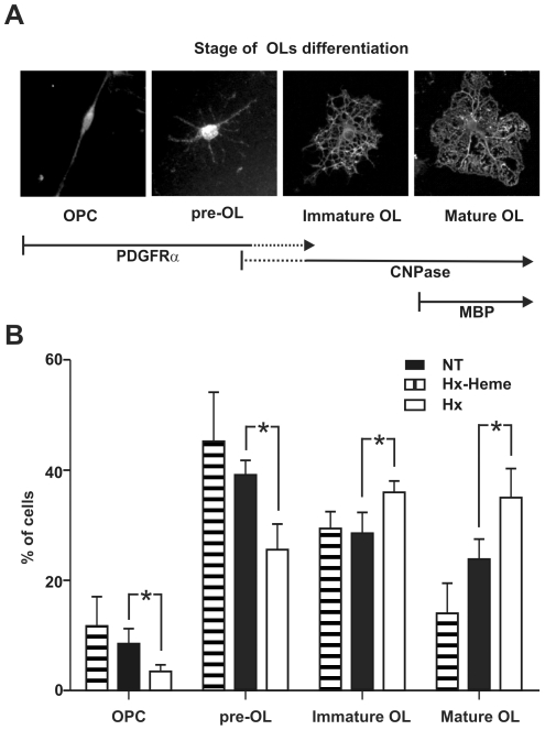
Myelin formation and maintenance are crucial for the proper function of the CNS and are orchestrated by a plethora of factors including growth factors, extracellular matrix components, metalloproteases and protease inhibitors. Hemopexin (Hx) is a plasma protein with high heme binding affinity, which is also locally produced in the CNS by ependymal cells, neurons and glial cells. We have recently reported that oligodendrocytes (OLs) are the type of cells in the brain that are most susceptible to lack of Hx, as the number of iron-overloaded OLs increases in Hx-null brain, leading to oxidative tissue damage. In the current study, we found that the expression of the Myelin Basic Protein along with the density of myelinated fibers in the basal ganglia and in the motor and somatosensory cortex of Hx-null mice were strongly reduced starting at 2 months and progressively decreased with age. Myelin abnormalities were confirmed by electron microscopy and, at the functional level, resulted in the inability of Hx-null mice to perform efficiently on the Rotarod. It is likely that the poor myelination in the brain of Hx-null mice was a consequence of defective maturation of OLs as we demonstrated that the number of mature OLs was significantly reduced in mutant mice whereas that of precursor cells was normal. Finally, in vitro experiments showed that Hx promotes OL differentiation. Thus, Hx may be considered a novel OL differentiation factor and the modulation of its expression in CNS may be an important factor in the pathogenesis of human neurodegenerative disorders.


Conflict of interest statement

Competing Interests: The authors have declared that no competing interests exist.

Figures

Figure 1
Figure 1. Reduction of MBP protein production in Hx−/− brain.
A) Western blot analysis of MBP expression in brain extracts of wild-type and Hx−/− mice. Cerebral cortex and basal ganglia region lysates were analyzed at two and twelve months of age. Representative experiments are shown. B) Band intensities were measured by densitometry and normalized to actin expression (AU: Arbitrary Unit). The overall MBP production was obtained by summing the relative intensities of the four isoforms recognized by the antibody (indicated by arrows in scanned gels). Densitometry data represent mean ± SEM; n = 3 for each genotype. *  =  P<0.05. Results shown are representative of three independent experiments.
Figure 2
Figure 2. The cortex of Hx−/− mice is hypomyelinated.
A) Coronal sections of a wild-type and a Hx−/− mouse at twelve months of age stained with Black-Gold reaction to detect myelinated fibers. Hx−/− mouse shows reduced myelination in cerebral cortex compared to wild-type (a, b) and the hypomyelination mainly affects the supragranular layers in motor and somatosensory cortex (arrows in c, d). Higher magnification shows that in layer I of Hx−/− mouse the staining is very weak compared to wild-type (e, f). Bar (c, d)  = 500 µm; Bar (e, f)  = 100 µm. B) Quantification of fiber density in motor cortical area, assessed at 2, 6 and 12 months of age, shows a severe reduction in Hx−/− mice. Data represent mean ± SEM, n = 3 mice for each genotype. *  = P<0.05, ***  = P<0.001.
Figure 3
Figure 3. Alteration of myelin ultrastructure in the absence of Hx.
EM analysis was performed on the corpus callosum of wild-type and Hx−/− mice at twelve month of age. A) Electron micrographs show that in Hx−/− mice the axons are hypomyelinated and the number of small myelinated axons is reduced in comparison to wild-types. Bar  = 1 µm. B) The distribution of myelin thickness in wild-type and Hx−/− mice fibers demonstrated that myelin sheath was thicker in Hx−/− fibers. P<0.0001. C) g-ratio scatter diagram in wild-type and Hx−/− mice fibers. Elevated g ratio values were observed for all axons in Hx−/− mice, indicating that impaired myelination affected axon of all sizes. P<0.001. D)The distribution of axonal size in wild-type and Hx−/− mice fibers showed that Hx−/− mice had bigger axons compared to controls. P<0.0001. n = 5 mice for each genotype.
Figure 4
Figure 4. Motor dysfunction in Hx−/− mice.
Accelerated Rotarod tests were performed every fifteen days from two to twelve months of age. The mean time score in which the mice walked in synchrony with the rod was recorded. Each test consisted of three consecutive trials. Hx−/− mice showed significant motor impairment starting from four months of age and increasing with age. Data represent mean ± SEM, n = 16 mice for each genotype. * =  P<0.05, **  = P<0.01, ***  = P<0.001.
Figure 5
Figure 5. Impaired OL development in Hx−/− mice.
Brain sections of wild-type and Hx−/− mice were immunoreacted to discriminate between OPCs and mature OLs and OPCs and OLs counted as reported in Materials and Methods. A) Quantification of PDGFRα-positive cells demonstrated similar numbers of OPCs in both cerebral cortex and corpus callosum in Hx−/− and wild-type mice at P10. On the contrary, the number of CC1-positive, GFAP-negative mature OLs in Hx−/− mice was strongly reduced compared to wild-type animals at P10 and P20. Data represent mean ± SEM, n = 3 mice for each genotype. **  = P<0.01, ***  = P<0.001. B) Maps, obtained with Neurolucida/Neuroexplorer, of brain sections of PDGFRα- (left) and CC1- (right) positive cells, respectively, in Hx−/− and wild-type mice at P10. Red  =  OPCs, blue  =  mature OLs. Note the reduced number of mature OLs in the supragranular layer of cortex in Hx−/− mice. C) Representative pictures of CC1/GFAP double staining for CC1 (brown) and GFAP (grey) in brain sections of a wild-type and a Hx−/− mouse. The latter shows a strong reduction in the number of CC1 positive cells in the cortex (arrows) and in corpus callosum, CC, (arrow-heads) compared to wild-type animal. Bar  = 50 µm.
Figure 6
Figure 6. Hx promotes OL differentiation.
OPCs were grown with or without Hx and the differentiation process was analyzed. A) Representative images showing the different developmental stages taken into consideration: stage I, OPCs (bipolar); stage II: pre-OL (primary branched); stage III: immature OL (secondary branched); stage IV: mature OL (secondary branched cells with membranous processes). Cells at stage I and II are PDGFRα positive, CNPase negative, cells at stage III are PDGFRα negative, CNPase positive and cells at stage IV are PDGFRα negative, CNPase and MBP positive. B) Kinetics of OL differentiation. Cells were cultured for 48 h in the absence (NT) or presence of Hx (Hx) or heme-Hx complex (Hx-heme), and the number of cells at each differentiation stage was counted as reported in Materials and Methods. Cells were scored by morphology and immunoreactivity to PDGFα and CNPase as shown in (A). Hx treatment accelerated the differentiation process whereas the heme-Hx complex was ineffective. *  =  P<0.05. Results shown are representative of three independent experiments.

References

    1. Bauer NG, ffrench-Constant C. Physical forces in myelination and repair: a question of balance? J Biol. 2009;8:78. - PMC - PubMed
    1. Bauer NG, Richter-Landsberg C, Ffrench-Constant C. Role of the oligodendroglial cytoskeleton in differentiation and myelination. Glia. 2009;57:1691–1705. - PubMed
    1. Baumann N, Pham-Dinh D. Biology of oligodendrocyte and myelin in the mammalian central nervous system. Physiol Rev. 2001;81:871–927. - PubMed
    1. Dangata YY, Kaufman MH. Myelinogenesis in the optic nerve of (C57BL x CBA) F1 hybrid mice: a morphometric analysis. Eur J Morphol. 1997;35:3–17. - PubMed
    1. Dyer CA. The structure and function of myelin: from inert membrane to perfusion pump. Neurochem Res. 2002;27:1279–1292. - PubMed

Publication types

MeSH terms