. 2011:17:1275-86.
Epub 2011 May 7.

Inhalative preconditioning with hydrogen sulfide attenuated apoptosis after retinal ischemia/reperfusion injury

Affiliations

Inhalative preconditioning with hydrogen sulfide attenuated apoptosis after retinal ischemia/reperfusion injury

Julia Biermann et al. Mol Vis. 2011.

Abstract

Purpose: Retinal ischemia/reperfusion (I/R) injury plays an important role in the pathophysiology of various ocular diseases. Retinal ganglion cells (RGCs) are particularly vulnerable to ischemia. Hydrogen sulfide (H(2)S) was recently shown to be neuroprotective in the brain and retina due to its antiapoptotic effects. Rapid preconditioning of retinal neurons by inhaled H(2)S before I/R injury may reduce apoptosis in the rat retina.

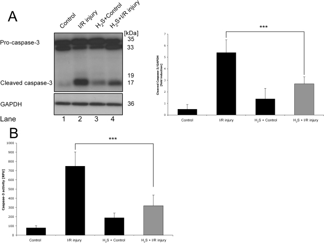
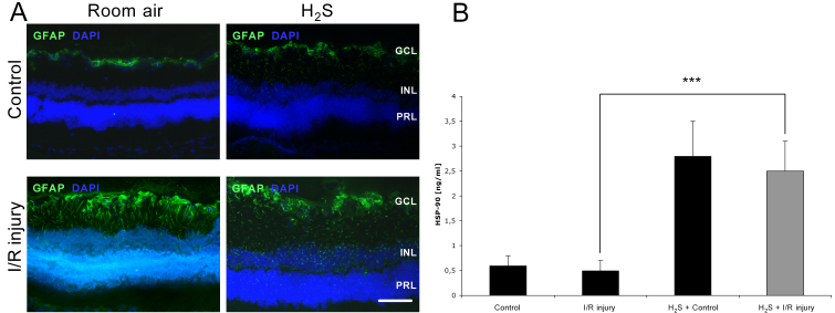
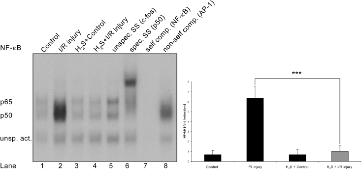
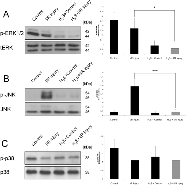
Methods: I/R injury was created on the left eye of rats (n=8) with or without inhaled H(2)S preconditioning (80 ppm) for one hour before ischemia. Densities of fluorogold prelabeled RGCs were analyzed 7 days after injury in retinal whole mounts. Retinal tissue was harvested to analyze protein expression of heat shock protein (HSP)-90 and the mitogen-activated protein kinases (MAPKs) c-jun N-terminal kinase (JNK), extracellular signal-regulated kinase (ERK)1/2 and p38 to elucidate a possible pathway of neuroprotection. DNA binding activity of the transcription factors nuclear factor-kappa-light-chain-enhancer of activated B-cells (NF-κB), cyclic adenosine monophosphate response element binding protein (CREB), and heat shock element (HSE), as well as caspase-3 cleavage and activity, were determined. Retinal sections were further assessed using anti-glial fibrillary acidic protein staining.

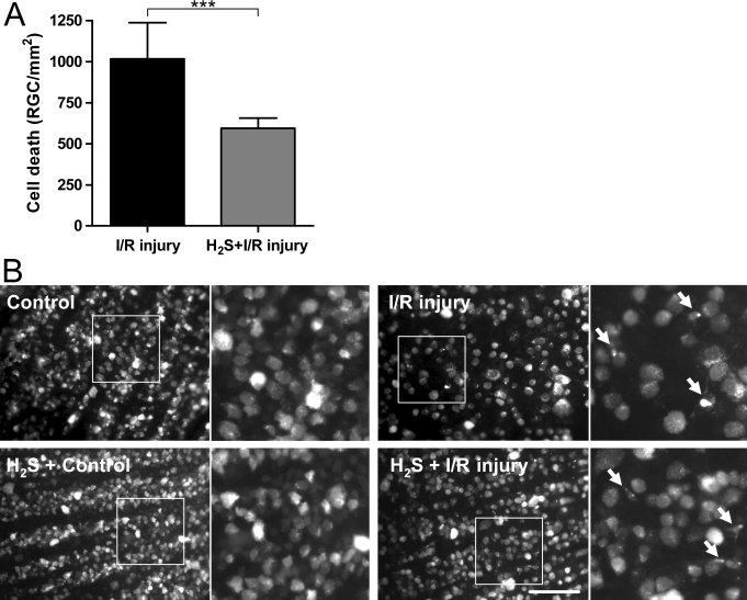
Results: RGC death after I/R injury decreased by 41.5% after H(2)S preconditioning compared to room air (p<0.001). H(2)S inhalation before ischemia reduced caspase-3 cleavage (p<0.001) and attenuated caspase-3 activity (p<0.001). Furthermore, HSP-90 expression was significantly elevated in the retina after H(2)S preconditioning. NF-κB but not CREB or HSE showed specific, H2S-dependent regulation, as well as the MAPKs ERK1/2 and JNK but not p38.

Conclusions: H(2)S preconditioning mediates antiapoptotic effects in retinal I/R injury, thus exhibiting neuroprotection. Based on these observations, H(2)S could represent a novel and promising therapeutic agent to counteract neuronal injuries in the eye. Further studies are needed to prove H(2)S's neuroprotective propensity using a postconditioning approach.


Figures

Figure 1
Figure 1
Attenuated retinal ganglion cell death after hydrogen sulfide preconditioning. A: Retinal ganglion cell (RGC) loss (RGC/mm2) presented as mean difference to individual control. Data are presented as mean±SD of eight experiments. Hydrogen sulfide (H2S) preconditioning decreased RGC death by 41.5% compared to room air seven days after ischemia/reperfusion (I/R) injury (***p<0.001). B: RGC densities were not significantly different in controls exposed to room air or H2S. Fluorogold (FG)-positive RGCs can be identified by morphological criteria (large round cell body, no processes, almost homogeneously labeled). In I/R treated eyes, many RGCs died and activated microglia cells (small cellular body, branching processes, inhomogeneously labeled; denoted with arrows in the extracts) stained FG-positive after RGC phagocytosis. H2S preconditioning partly antagonized this effect, leading to significantly higher cell densities. The scale bar represents 100 µm.
Figure 2
Figure 2
Hydrogen sulfide preconditioning–mediated antiapoptotic effects. A: Effect of hydrogen sulfide (H2S) preconditioning on caspase-3 activation 24 h after unilateral ischemia. Pro-caspase-3 and caspase-3 levels were determined using specific antibodies. Histograms represent the densitometric ratio of caspase-3 cleavage compared with loading control (glyceraldehyde 3-phosphate dehydrogenase [GAPDH]). The amount of pro-caspase-3 and protein loading seemed comparable in all groups (lanes 1–4). Compared to control, ischemia/reperfusion (I/R) injury led to a significant cleavage from pro-caspase-3 to active caspase-3 (lane 1 versus 2). H2S preconditioning before I/R injury significantly reduced cleavage of pro-caspase-3 to caspase 3 (lane 4 versus 2; ***p<0.001). Data are presented as mean±SD of five experiments. B: Fluorogenic caspase-3 assay (DEVDase assay) of full retinal protein lysates 24 h after I/R injury. Caspase-3 activity was low in control eyes (room air) and was not significantly affected by H2S inhalation in controls. I/R injury increased the activity (p<0.001 compared with control eye). In contrast, preconditioning with inhaled H2S significantly reduced caspase-3 activity in ischemic tissue. Results are given in RFUs. Data are presented as mean±SD of eight experiments. ***p<0.001 I/R injury versus H2S + I/R injury.
Figure 3
Figure 3
Hydrogen sulfide–attenuated glial fibrillary acidic protein expression and increased heat shock protein (HSP)-90 expression in retinal tissue. Effect of hydrogen sulfide (H2S) preconditioning on retinal glial fibrillary acidic protein (GFAP; A) and HSP-90 (B) expression. A: Cross-sections of the retinas 7 days after unilateral ischemia/reperfusion (I/R) injury. In controls with and without H2S preconditioning, GFAP was only positive in Müller cells and astrocytes in the ganglion cell layer (GCL). After ischemia, GFAP was upregulated in Müller cells. Their processes, extending through all retinal layers, became strongly GFAP positive. This upregulation seemed to be stronger in the I/R injury + room air group. The scale bar represents 50 µm. B: In nonpreconditioned eyes (control and ischemic), retinal HSP-90 expression remained at the baseline level. H2S preconditioning in control eyes significantly increased HSP-90 expression. H2S inhalation before I/R injury significantly induced retinal HSP-90 expression compared with I/R injury alone (***p<0.001). Data are presented as mean±SD of eight experiments.
Figure 4
Figure 4
Hydrogen sulfide counteracted ischemia/reperfusion-induced DNA-binding activity of nuclear factor-kappaB . Effect of hydrogen sulfide (H2S) on the DNA-binding activity of nuclear factor-kappaB (NF-κB) in retinal tissue 24 h after unilateral ischemia. Control eyes with and without H2S preconditioning did not reveal any DNA binding of NF-κB (lanes 1, 3). Compared to control, ischemia/reperfusion (I/R) injury increased the DNA binding of NF-κB significantly (lane 1 versus 2; sixfold induction versus control, p<0.001). H2S inhalation before I/R injury counteracted the ischemia-induced DNA binding of NF-κB completely (lane 4 versus 2, 5.8-fold reduction versus I/R injury, ***p<0.001). Specific supershift analysis revealed that p50 is the main part of the transactive NF-κB domain (lane 6), while the unspecific antibody (c-fos [AP-1]) did not lead to a supershift. The oligo’s sensitivity was demonstrated by competition experiments with unlabeled NF-κB (lane 7) and unlabeled AP-1 (lane 8). The histogram represents the densitometric ratio of NF-κB compared with the control group. Data are presented as mean±SD of five experiments.
Figure 5
Figure 5
Hydrogen sulfide–induced differential mitogen-activated protein kinase regulation in the retina. Effect of hydrogen sulfide (H2S) preconditioning on mitogen-activated protein kinases (MAPKs) phosphorylated (p)- extracellular signal-regulated kinase (ERK)1/2 (A), p- c-jun N-terminal kinase (JNK; B), and p-p38 (C) 24 h after unilateral ischemia. MAPK levels were determined using specific antibodies. The histograms represent the densitometric ratio of MAPKs compared with its nonphosphorylated form, total (t)ERK1/2, JNK, or p38. A: p-ERK1/2 is suppressed through inhalational H2S preconditioning. While p-ERK was baseline activated in the nonpretreated animals (control and ischemia/reperfusion [I/R] injury), H2S significantly inhibited ERK1/2 phosphorylation in both control and ischemic retinas (fourfold reduction in H2S + I/R injury versus I/R injury; *p<0.05). B: I/R injury per se significantly increased the phosphorylation of the JNK (3.8-fold induction in I/R injury versus control; p<0.001). Again, preconditioning with inhalative H2S inhibited JNK phosphorylation completely (3.7-fold reduction in H2S + I/R injury versus I/R injury; ***p<0.001). C: The phosphorylation of p38 MAPK was comparable in all groups without significant differences. Data are presented as mean±SD of eight experiments.

References

    1. Verma D. Pathogenesis of diabetic retinopathy–the missing link? Med Hypotheses. 1993;41:205–10. - PubMed
    1. Archer DB. Tributary vein obstruction: pathogenesis and treatment of sequelae. Doc Ophthalmol. 1976;40:339–60. - PubMed
    1. Hayreh SS. Ischemic optic neuropathy. Int Ophthalmol. 1978;1:9–18. - PubMed
    1. Nickells RW. Retinal ganglion cell death in glaucoma: the how, the why, and the maybe. J Glaucoma. 1996;5:345–56. - PubMed
    1. Schmidt KG, Pillunat LE, Osborne NN. Ischemia and hypoxia. An attempt to explain the different rates of retinal ganglion cell death in glaucoma. Ophthalmologe. 2004;101:1071–5. - PubMed

MeSH terms

LinkOut - more resources