Review
. 2011 Aug;20(8):1298-345.
doi: 10.1002/pro.666.

The N-end rule pathway and regulation by proteolysis

Affiliations
Review

The N-end rule pathway and regulation by proteolysis

Alexander Varshavsky. Protein Sci. 2011 Aug.

Abstract

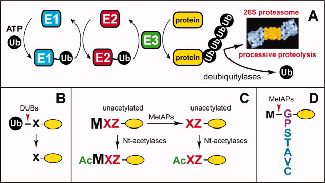
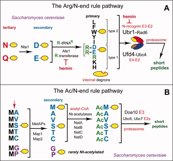
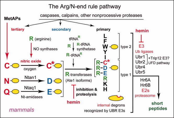
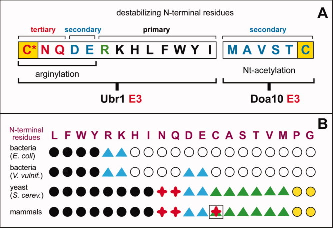
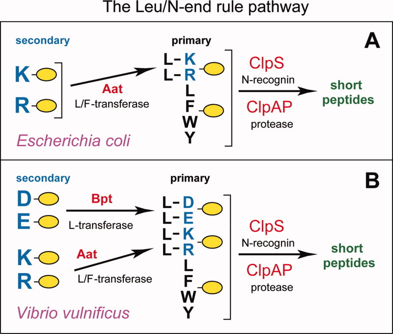
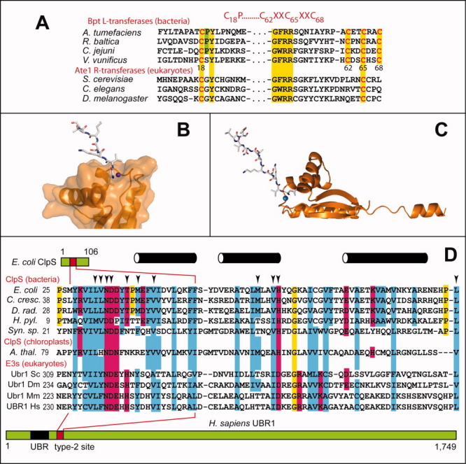
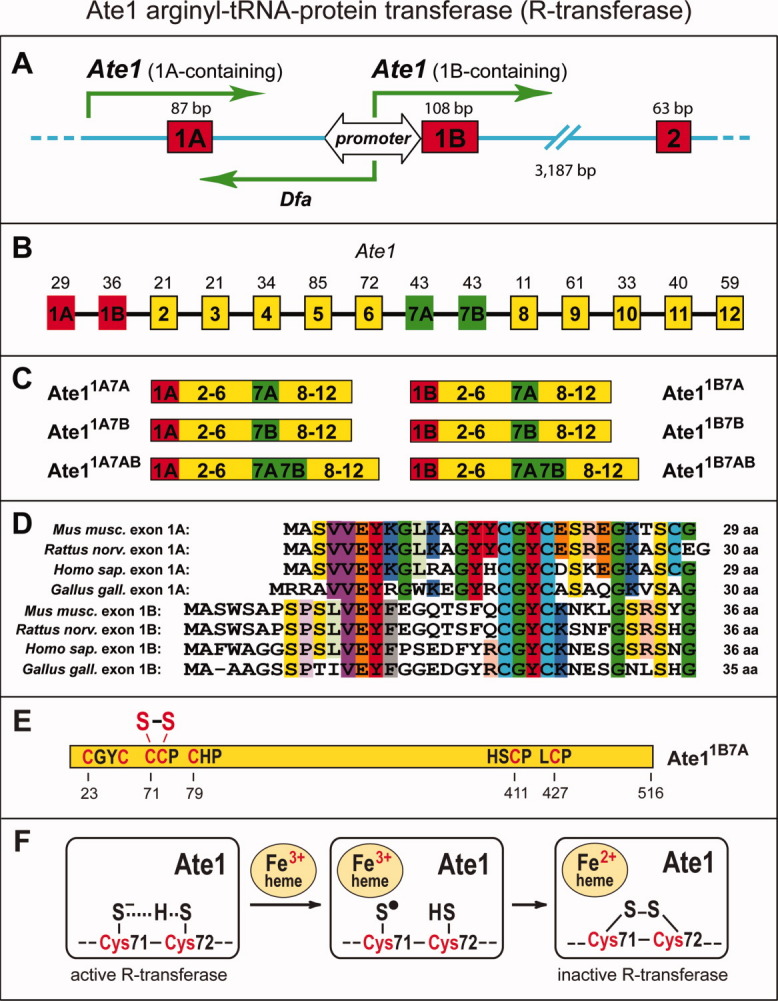
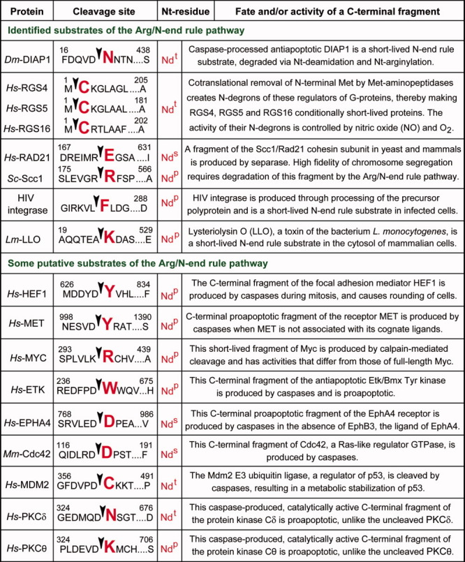
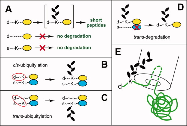
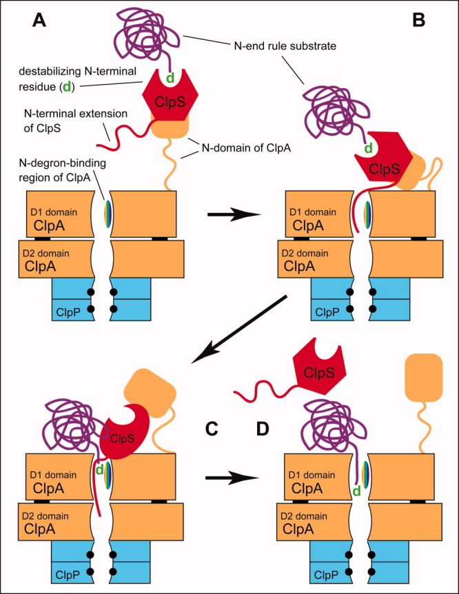
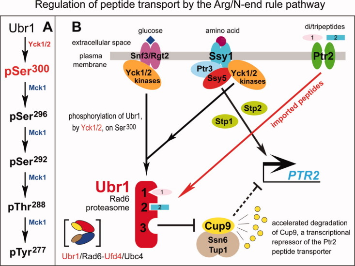
The N-end rule relates the regulation of the in vivo half-life of a protein to the identity of its N-terminal residue. Degradation signals (degrons) that are targeted by the N-end rule pathway include a set called N-degrons. The main determinant of an N-degron is a destabilizing N-terminal residue of a protein. In eukaryotes, the N-end rule pathway is a part of the ubiquitin system and consists of two branches, the Ac/N-end rule and the Arg/N-end rule pathways. The Ac/N-end rule pathway targets proteins containing N(α) -terminally acetylated (Nt-acetylated) residues. The Arg/N-end rule pathway recognizes unacetylated N-terminal residues and involves N-terminal arginylation. Together, these branches target for degradation a majority of cellular proteins. For example, more than 80% of human proteins are cotranslationally Nt-acetylated. Thus most proteins harbor a specific degradation signal, termed (Ac)N-degron, from the moment of their birth. Specific N-end rule pathways are also present in prokaryotes and in mitochondria. Enzymes that produce N-degrons include methionine-aminopeptidases, caspases, calpains, Nt-acetylases, Nt-amidases, arginyl-transferases and leucyl-transferases. Regulated degradation of specific proteins by the N-end rule pathway mediates a legion of physiological functions, including the sensing of heme, oxygen, and nitric oxide; selective elimination of misfolded proteins; the regulation of DNA repair, segregation and condensation; the signaling by G proteins; the regulation of peptide import, fat metabolism, viral and bacterial infections, apoptosis, meiosis, spermatogenesis, neurogenesis, and cardiovascular development; and the functioning of adult organs, including the pancreas and the brain. Discovered 25 years ago, this pathway continues to be a fount of biological insights.


Figures

Figure 1
Figure 1
The ubiquitin-proteasome system, the ubiquitin fusion technique, and N-terminal processing of newly formed proteins. A: The ubiquitin-proteasome system (Ub system).,– The conjugation of Ub to other proteins involves a preliminary ATP-dependent step in which the last residue of Ub (Gly76) is joined, via a thioester bond, to a Cys residue of the E1 (Ub-activating) enzyme. The “activated” Ub moiety is transferred to a Cys residue in one of several Ub-conjugating (E2) enzymes, and from there, through an isopeptide bond, to a Lys residue of an ultimate acceptor, denoted as “protein”. E2 enzymes function as subunits of E2-E3 Ub ligase complexes that can produce substrate-linked poly-Ub chains. Such chains have specific Ub-Ub topologies, depending on the identity of a Lys residue of Ub (which contains several lysines) that forms an isopeptide bond with C-terminal Gly of the adjacent Ub moiety in a chain. Specific poly-Ub chains can confer the degradation of a substrate by the 26S proteasome or other metabolic fates. Monoubiquitylation of some protein substrates can also occur, and has specific functions. One role of E3 is the recognition of a substrate's degradation signal (degron). Individual mammalian genomes encode at least a 1,000 distinct E3 Ub ligases. B: The Ub fusion technique., In eukaryotes, linear fusions of Ub to other proteins are cotranslationally cleaved by deubiquitylases at the last residue of Ub, making it possible to produce, in vivo, different residues at the N-termini of otherwise identical proteins. C: N-terminal processing of nascent proteins by Nα-terminal acetylases (Nt-acetylases) and Met-aminopeptidases (MetAPs). “Ac” denotes the Nα-terminal acetyl moiety. M, Met. X and Z, single-letter abbreviations for any amino acid residue. Yellow ovals denote the rest of a protein. D: Met-aminopeptidases (MetAPs) cleave off the N-terminal Met residue if a residue at Position 2 belongs to the set of residues shown. Gly and Pro at Position 2 are depicted in a different color because these residues, in contrast to other small residues, are rarely Nt-acetylated after the removal of N-terminal Met [Fig. 2(B)]. [Color figure can be viewed in the online issue, which is available at wileyonlinelibrary.com.]
Figure 2
Figure 2
The N-end rule pathway in Saccharomyces cerevisiae. A: The Arg/N-end rule pathway. See the main text for details. Yellow ovals denote the rest of a protein substrate. “Primary”, “secondary” and “tertiary” denote mechanistically distinct subsets of destabilizing N-terminal residues. The physically associated Ubr1 (N-recognin) and Ufd4 E3s have substrate-binding sites that recognize internal (non-N-terminal) degrons in substrates of the Arg/N-end rule pathway that lack N-degrons. Ubr1 (but not Ufd4) recognizes N-degrons as well. B: The Ac/N-end rule pathway. Red arrow on the left indicates the removal of N-terminal Met by Met-aminopeptidases (MetAPs). This Met residue is retained if a residue at Position 2 is nonpermissive (too large) for Met-aminopeptidases [Fig. 1(D)]. If the (retained) N-terminal Met or N-terminal Ala, Val, Ser, Thr and Cys are followed by residues that allow Nt-acetylation (see the main text), these N-terminal residues are usually Nt-acetylated.– The resulting N-degrons are called AcN-degrons. The term “secondary” refers to the necessity of modification (Nt-acetylation) of a destabilizing N-terminal residue before a protein can be recognized by a cognate Ub ligase. Proteins containing AcN-degrons are targeted for ubiquitylation and proteasome-mediated degradation by the Doa10 E3 N-recognin, in conjunction with the Ubc6 and Ubc7 E2 enzymes. Although Gly and Pro can be made N-terminal by MetAPs, and although Doa10 can recognize Nt-acetylated Gly and Pro, few proteins with N-terminal Gly or Pro are Nt-acetylated.– [Color figure can be viewed in the online issue, which is available at wileyonlinelibrary.com.]
Figure 3
Figure 3
The mammalian Arg/N-end rule pathway. See the main text for details. N-terminal residues are indicated by single-letter abbreviations for amino acids. Yellow ovals denote the rest of a protein substrate. “Primary”, “secondary” and “tertiary” denote mechanistically distinct subsets of destabilizing N-terminal residues. C* denotes oxidized N-terminal Cys, either Cys-sulfinate or Cys-sulfonate, produced in vivo through reactions that require both nitric oxide (NO) and oxygen., The mammalian N-recognins Ubr1, Ubr2, Ubr4, and Ubr5 (Edd) have multiple substrate binding sites that also recognize internal (non-N-terminal) degrons in other substrates of the Arg/N-end rule pathway, the ones that lack N-degrons. A question mark after Trip12 (which mediates the mammalian UFD pathway and is a sequelog of the S. cerevisiae Ufd4 E3) denotes the untested possibility that mammalian Ubr1 and/or Ubr2 form complexes with Trip12, by analogy with the Ubr1–Ufd4 complex in S. cerevisiae [Fig. 2(A)]. [Color figure can be viewed in the online issue, which is available at wileyonlinelibrary.com.]
Figure 4
Figure 4
Rule books of N-end rules. A: The N-end rule in eukaryotes. It results from combined activities of the Arg/N-end rule and Ac/N-end rule pathways. In eukaryotes that produce NO, the N-terminal Cys residue (in yellow rectangles) can be targeted, alternatively, by either one of the two branches of the N-end rule pathway, with oxidized Cys marked by an asterisk (see Overview of the N-End Rule Pathway section). B: A comparison of rule books of N-end rule pathways in different organisms, indicated on the left. Black circles, blue or green triangles, and red crosses denote primary (Ndp), secondary (Nds) and tertiary (Ndt) destabilizing N-terminal residues, respectively. Blue triangles denote secondary destabilizing N-terminal residues that involve either Nt-leucylation (in bacteria) or Nt-arginylation (in eukaryotes). Green triangles denote secondary destabilizing N-terminal residues that involve Nt-acetylation. N-terminal Cys is denoted by both a green triangle and a red cross, given its alternative functioning as a part of NO/O2-mediated N-degrons or AcN-degrons. Open circles, in bacterial N-end rules, denote stabilizing (nondestabilizing) N-terminal residues. Yellow circles, in eukaryotic N-end rules, denote Pro and Gly. These N-terminal residues are rarely Nt-acetylated and therefore, operationally, are stabilizing (nondestabilizing) residues. But in some proteins with N-terminal Pro or Gly these residues can be Nt-acetylated. If other components of an AcN-degron are also in place (see The Ac/N-End Rule Pathway section), such proteins can become substrates of the Ac/N-end rule pathway. [Color figure can be viewed in the online issue, which is available at wileyonlinelibrary.com.]
Figure 5
Figure 5
Bacterial N-end rule pathways. A: The E. coli Leu/N-end rule pathway.,, See Overview of the N-End Rule Pathway, Structure and Targeting of N-degrons, and Prokaryotic N-End Rule Pathways sections for details. The Aat L/F-transferase conjugates (largely) Leu to N-terminal Arg or Lys. N-end rule substrates bearing primary (bulky hydrophobic) destabilizing N-terminal residues are recognized by the ClpS N-recognin and are delivered for degradation to the ClpAP protease. B: The Leu/N-end rule pathway in another gram-negative bacterium, V. vulnificus, which contains both the Aat L/F-transferase and the Bpt L-transferase. As a result, N-terminal Asp and Glu, which are stabilizing (nondestabilizing) residues in E. coli, are secondary destabilizing residues in the V. vulnificus Leu/N-end rule pathway. [Color figure can be viewed in the online issue, which is available at wileyonlinelibrary.com.]
Figure 6
Figure 6
Bpt L-transferases and ClpS N-recognins. A: Sequence alignments of bacterial Bpt L-transferases and eukaryotic Ate1 R-transferases. The sequelogy (sequence similarity1) between Bpt and Ate1 encompasses more of their sequences than shown here. Ate1 R-transferases lack a sequence motif (its consensus, in red, is shown at the top of the diagram) that Bpt L-transferases uniformly contain. This motif is characteristic of proteins that bind to a Fe-S cluster, (Prokaryotic N-End Rule Pathways section). B: Surface representation of the C-terminal domain of the E. coli ClpS N-recognin in a complex with an 11-mer peptide (shown as a stick model) that bears N-terminal Leu, a primary destabilizing (Ndp) residue in the Leu/N-end rule pathway. Blue sphere, water molecule. C: Ribbon representation of the full-length 12-kDa ClpS in the same complex. D: Sequence alignments of the ∼70-residue domain of bacterial ClpS N-recognins in Caulobacter crescentus, E. coli, Deinococcus radiodurans, Helicobacter pylori, Synechocystis sp. PCC6803 (the latter a photosynthesis-capable cyanobacterium), and in chloroplast (A. thaliana). This region of ClpS binds to N-terminal Ndp residues of the Leu/N-rule pathway. ClpS sequences are aligned with sequelogous regions of eukaryotic (S. cerevisiae, D. melanogaster, M. musculus, H. sapiens) Ubr1 N-recognins (they are ∼20-fold larger than ClpS) that encompass the Type-2 substrate-binding site of Ubr1 [Fig. 7(A)]. The specificity of this Ubr1 binding site for bulky hydrophobic N-terminal Ndp residues is nearly the same as the specificity of ClpS, except that the Ubr1 site binds to N-terminal Ile as well [Fig. 2(A)], in contrast to ClpS (see the main text). Arrowheads indicate the positions of crystallographically determined contacts between the ClpS of C. crescentus and an N-end rule peptide. Black cylinders indicate α-helices in this region of ClpS. [Color figure can be viewed in the online issue, which is available at wileyonlinelibrary.com.]
Figure 7
Figure 7
Structural organization and phosphorylation of the Ubr1 N-recognin. A: Phosphorylated residues of the S. cerevisiae Ubr1 E3 N-recognin of the Arg/N-end rule pathway are indicated above the diagram. The regions containing the Type-1 substrate-binding site (UBR domain), the Type-2 substrate-binding site, the BRR (basic residues-rich) domain, the Cys/His-rich RING domain and the AI (autoinhibitory) domain,,,,– are also indicated. B: The “primed” cascade of Ubr1 phosphorylation. The initial phosphorylation of Ubr1 on Ser by the Yck1/Yck2 kinases of the casein kinase type-I family makes possible (primes) the subsequent, apparently sequential phosphorylation of Ubr1 by Mck1, a Gsk3-type kinase, on Ser, Ser, Thr, and Tyr. Also indicated is the identified function of the Ser phosphorylation of Ubr1 in the control of peptide import. [Color figure can be viewed in the online issue, which is available at wileyonlinelibrary.com.]
Figure 8
Figure 8
UBR domains in N-recognins and putative N-recognins of the mammalian Arg/N-end rule pathway. A: Ribbon diagram of the ∼80-residue S. cerevisiae UBR domain (Fig. 7A) in the complex with the RLGES peptide that bears N-terminal Arg, a Type-1 Ndp residue. The bound RLGES is shown as a stick model, with carbon atoms colored yellow. Several residues are marked with a black sphere and numbered to facilitate the tracing of the polypeptide chain. The names of residues of the RLGES peptide are in red, with the letter “s” (substrate) appended to their position numbers. Side chains of residues in the UBR domain that are present near missense mutations in UBR1 of patients with Johanson–Blizzard syndrome (JBS; C.-S. Hwang et al., unpublished data) are shown in a stick form, with carbon atoms colored green. Three coordinated zinc ions of the UBR domain are shown as red spheres. B: Molecular surface of the S. cerevisiae UBR domain. Negatively and positively charged surfaces are shaded red and blue, respectively. The bound RLGES peptide is shown in yellow. Some residues of Ubr1 that comprise the N-degron-binding cleft are labeled. C: Diagram of the mammalian UBR-domain family of E3 Ub ligases, showing both UBR and other domains of these E3s (RING, HECT, PHD, CRD and F-box) that contribute to recognition and ubiquitylation of protein substrates., Ubr1, Ubr2, Ubr4, and Ubr5/Edd of this set are operationally defined N-recognins of the mammalian Arg/N-end rule pathway in that they specifically bind to the Type-1 and/or Type-2 destabilizing N-terminal residues, whereas Ubr3, Ubr6, and Ubr7 are not N-recognins, (The double-E3 design of the Arg/N-end rule pathway section). [Color figure can be viewed in the online issue, which is available at wileyonlinelibrary.com.]
Figure 9
Figure 9
Splicing-derived isoforms of the Ate1-encoded Arg-tRNA-protein transferase (R-transferase) and its inhibition by hemin. A: The bidirectional DfaPAte1 promoter (containing a CpG island) upstream of exon 1B of the mouse Ate1 gene., Green arrows indicate transcriptional units oriented in both directions from DfaPAte1, and also from an unmapped “upstream” promoter that mediates the expression of Ate1 transcripts containing exon 1A. The locations and sizes of some Ate1 exons are shown as well. B: The exons, including alternative exons, of the mouse Ate1 gene, with deduced lengths of the corresponding polypeptide segments indicated on top. C: Mouse R-transferase isoforms (and their designations) that are produced through alternative splicing of Ate1 pre-mRNA. D: Sequence comparisons of translated vertebrate Ate1 exons 1A amongst themselves and with the set of longer but also sequelogous alternative exons 1B. Most of recurrent amino acid identities are highlighted by color. Mus musculus, mouse; Rattus norvegicus, rat; Homo sapiens, human; Gallus gallus, chicken. E: The mouse ATE11B7A isoform, with locations of significant Cys-containing motifs, including the vicinal Cys and Cys residues. A disulfide bond between them is the result of hemin-mediated oxidation and functional inactivation of R-transferase. F: Diagram of the previously proposed redox mechanism of the hemin-mediated disulfide formation between Cys and Cys of Ate1. [Color figure can be viewed in the online issue, which is available at wileyonlinelibrary.com.]
Figure 10
Figure 10
Confirmed and putative N-end rule substrates produced by caspases and other nonprocessive proteases. Amino acid residues are indicated by single-letter abbreviations. Arrowheads and enlarged residues, in red, indicate the cleavage sites and N-terminal residues of the corresponding C-terminal fragments. A number on the left represents the first residue of a protein (numbered as in the full-length protein) that is shown in the diagram. A number on the right represents the last residue of a full-length protein. The prefixes Dm, Hs, Mm, and Sc refer to proteins of D. melanogaster, H. sapiens, M. musculus and S. cerevisiae, respectively. See Substrates of the N-End Rule Pathway section for a description of specific protein fragments cited in this list. [Color figure can be viewed in the online issue, which is available at wileyonlinelibrary.com.]
Figure 11
Figure 11
Organization and cis-trans targeting of eukaryotic N-degrons. A: Three determinants of N-degron. d, a destabilizing N-terminal residue. K, a “ubiquitylatable” internal Lys residue. The absence of one of these determinants abrogates polyubiquitylation of a protein, despite the presence of another determinant. The third determinant of N-degron is an unstructured region that is required for polyubiquitylation and/or the initiation of degradation of a polyubiquitylated N-end rule substrate by the 26S proteasome. See Substrates of the N-End Rule Pathway section for references and details. B and C: cis versus trans polyubiquitylation of an oligomeric N-end rule substrate that results in the degradation of a subunit that becomes linked to a poly-Ub chain (see Substrates of the N-End Rule Pathway section). D: trans-degradation, in which a specific subunit of oligomeric protein is polyubiquitylated but is not degraded by the 26 proteasome, for example, because it lacks an unstructured region that is required for the initiation of degradation. Instead, a subunit-selective degradation of another, nonubiquitylated subunit takes place. This mode of degradation was demonstrated by the Matouschek laboratory for oligomeric substrates of the UFD pathway. It remains to be determined whether the analogous (hypothetical) trans-degradation of an oligomeric N-end rule substrate can also occur. E: The 1989–1996 hairpin insertion model of protein targeting by the 26S proteasome. No details of the 26S proteasome structure (such as the 19S regulatory particle (RP)) are shown in this 1996 diagram, and the sizes of specific components such as Ub moieties, the poly-Ub chain and the proteasome, are not to scale (see Substrates of the N-End Rule Pathway section). [Color figure can be viewed in the online issue, which is available at wileyonlinelibrary.com.]
Figure 12
Figure 12
Targeting and degradation of N-end rule substrates by the bacterial Leu/N-end rule pathway. This model, proposed by Román-Hernandez et al. and based on studies by the Baker, Sauer, Bukau, Maurizi, and other laboratories,,,,,, is described in the main text. A folded polypeptide chain of an N-end rule substrate (in purple color) is depicted “explicitly”, in contrast to solid-body renderings of ClpS and ClpAP. Black circles in the ClpP moiety indicate its proteolytic active sites. See Substrates of the N-End Rule Pathway section for details. [Color figure can be viewed in the online issue, which is available at wileyonlinelibrary.com.]
Figure 13
Figure 13
Steric shielding of the Nt-acetylated N-terminal residue of a subunit in a protein complex. Shown here is a part of the crystal structure, by the Barford laboratory, of a complex between the Hcn1 and Cut9 subunits of the S. pombe APC/C Ub ligase. In this structure, the Nt-acetylated N-terminal Met residue of Hcn1 is enclosed within a chamber formed by the Cut9 subunit, including its interface with the other Cut9 subunit in the heterotetramer of Hcn1 and Cut9. N-terminal region of Hcn1 is shown in cyan as a stick model, and Cut9 is depicted as a cut-out surface representation, to show the chamber's interior. [Color figure can be viewed in the online issue, which is available at wileyonlinelibrary.com.]
Figure 14
Figure 14
Spalogy (spatial similarity1) between the Ntaq1 NtQ-amidase and Factor XIII transglutaminase. A and B: Crystal structures of the human Ntaq1 (C8orf32) NtQ-amidase (PDB: 3C9Q) and FXIII transglutaminase (PDB: 1FIE), respectively. C8orf32 is an initially uncharacterized human protein the structure of which was deposited in the Protein Data Bank (PDB: 3C9Q) by the Center for Eukaryotic Structural Genomics, and was later shown, by Wang et al., to be the Ntaq1 NtQ-amidase. C and D: Structures around the active sites of Ntaq1 NtQ-amidase and Factor XIII transglutaminase, respectively. These regions are circled in A and B. C and D: The catalytic triad (Cys, His,373 and Asp396) of Factor XIII transglutaminase (D; ref. 341) and the corresponding residues (Cys, His, and Asp97) of human Ntaq1 (C) are indicated. Despite the striking spalogy between these regions of two enzymes (C and D), there is no significant sequelogy (sequence similarity) between them. [Color figure can be viewed in the online issue, which is available at wileyonlinelibrary.com.]
Figure 15
Figure 15
Regulation of peptide import by the Arg/N-end rule pathway in S. cerevisiae, and inputs by the amino acid-sensing SPS pathway. A: The “primed” cascade of Ubr1 phosphorylation in which the Yck1/Yck2-mediated phosphorylation on Ser of Ubr1 is essential for the normal regulation of peptide import [see also the legend to Fig. 7(A,B)]. B: Ubr1-mediated regulation of peptide import, and the involvement of the SPS pathway.,,,, Cup9 is a transcriptional repressor of the regulon that includes PTR2, which encodes the major importer of di/tripeptides. In the absence of Ubr1 (in ubr1Δ cells), Cup9 becomes long-lived, accumulates to high levels, and extinguishes expression of Ptr2. Therefore, ubr1Δ cells cannot import di/tripeptides. In wild-type (UBR1) cells growing in the absence of extracellular di/tripeptides, a relatively low but nonzero number of Ubr1 molecules have their third substrate-binding site “open” (not autoinhibited) and therefore can target Cup9 for degradation (t1/2 ∼ 5 min) via its internal degron, resulting in a low but significant steady-state concentration of Cup9 and, thus, a weak but significant expression of the Ptr2 transporter. In wild-type (UBR1) cells growing in the presence of extracellular di/tripeptides (some of which bear Type-1 and Type-2 destabilizing N-terminal residues), the imported peptides interact with the Type-1 and Type-2 binding sites of Ubr1. This binding allosterically increases the fraction of Ubr1 molecules whose third (Cup9-specific) site is “open” (active). The resulting decrease in the half-life of Cup9 (from ∼5 min to below 1 min) results in a low concentration of Cup9, and consequently to a strong induction of the Ptr2 transporter.,, Also shown is the amino acid-sensing SPS pathway (see Functions of the N-end rule pathway vis-á-vis their mechanisms section for details and additional references), which can influence the import of peptides at least in part through the Yck1/Yck2-mediated phosphorylation of Ubr1 on Ser. This phosphorylation is required (through a mechanism that remains to be determined) for normal levels of Ubr1 activity in the Ptr2-Cup9-Ubr1 circuit. [Color figure can be viewed in the online issue, which is available at wileyonlinelibrary.com.]

References

    1. Varshavsky A. Spalog and sequelog: neutral terms for spatial and sequence similarity. Curr Biol. 2004;14:R181–R183. - PubMed
    1. Varshavsky A. Naming a targeting signal. Cell. 1991;64:13–15. - PubMed
    1. Hwang C-S, Shemorry A, Varshavsky A. N-terminal acetylation of cellular proteins creates specific degradation signals. Science. 2010;327:973–977. - PMC - PubMed
    1. Bachmair A, Finley D, Varshavsky A. In vivo half-life of a protein is a function of its amino-terminal residue. Science. 1986;234:179–186. - PubMed
    1. Varshavsky A. The N-end rule: functions, mysteries, uses. Proc Natl Acad Sci USA. 1996;93:12142–12149. - PMC - PubMed

Publication types