

Telomere length in myelodysplastic syndromes
- PMID: 21635204
- PMCID: PMC4350661
- DOI: 10.3109/10428194.2011.568648
Telomere length in myelodysplastic syndromes
Abstract
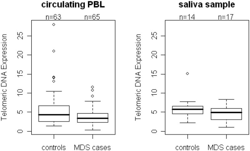
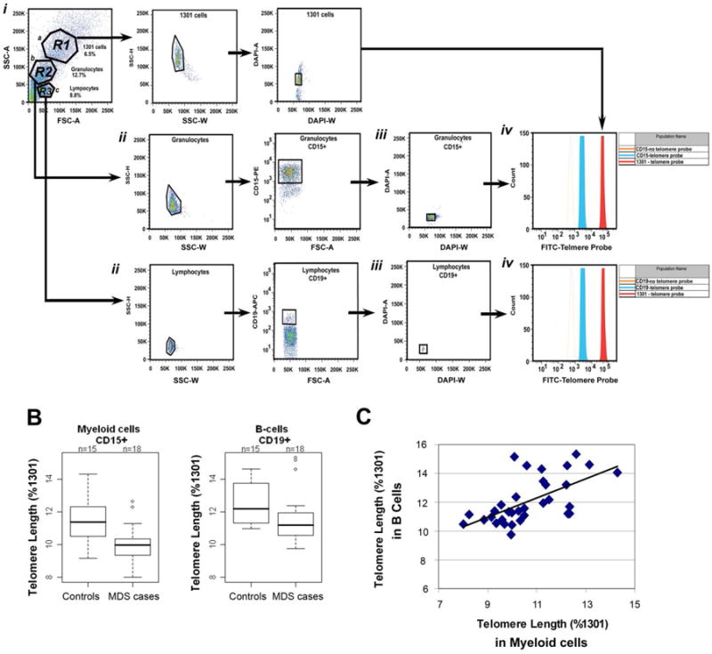
The relationship between telomere length (TL) and predisposition to myelodysplastic syndromes (MDS) remains unclear. We compared peripheral blood leukocyte (PBL) TL among cases of histologically confirmed MDS (n = 65) who were treatment-naive with no prior cancer history to age-matched controls (n = 63). Relative TL was measured in PBLs and saliva by quantitative polymerase chain reaction (PCR) and in CD15+ and CD19+ cells by flow cytometry-fluorescence in situ hybridization (flow-FISH). Human telomerase reverse transcriptase gene (hTERT) mutations were assessed by PCR. After adjustment for age and sex, relative TLs were reduced in PBLs (p = 0.02), CD15+ (p = 0.01), CD19+ (p = 0.25), and saliva (p = 0.13) in MDS cases versus controls, although only the PBL and CD15+ results were statistically significant. Among MDS cases, CD15+ and CD19+ cell TLs were positively correlated (p = 0.03). PBL TL was reduced among those occupationally exposed to paints and pesticides, but was not associated with hTERT genotype. Future studies are needed to further investigate constitutional telomere attrition as a possible predisposing factor for MDS.
Figures
References
-
- Baird DM, Kipling D. The extent and significance of telomere loss with age. Ann N Y Acad Sci. 2004 Jun;1019:265–8. - PubMed
-
- Londono-Vallejo JA. Telomere length heterogeneity and chromosome instability. Cancer Lett. 2004 Aug 30;212(2):135–44. - PubMed
-
- Boultwood J, Fidler C, Kusec R, et al. Telomere length in myelodysplastic syndromes. Am J Hematol. 1997 Dec;56(4):266–71. - PubMed
-
- Brummendorf TH, Holyoake TL, Rufer N, et al. Prognostic implications of differences in telomere length between normal and malignant cells from patients with chronic myeloid leukemia measured by flow cytometry. Blood. 2000 Mar 15;95(6):1883–90. - PubMed
-
- Ohyashiki JH, Iwama H, Yahata N, et al. Telomere stability is frequently impaired in high-risk groups of patients with myelodysplastic syndromes. Clin Cancer Res. 1999 May;5(5):1155–60. - PubMed
Publication types
MeSH terms
Substances
Grants and funding
LinkOut - more resources
Full Text Sources
Other Literature Sources
Medical
Research Materials
Miscellaneous