. 2011 Aug;133(4):452-68.
doi: 10.1111/j.1365-2567.2011.03456.x. Epub 2011 Jun 3.

T regulatory cells participate in the control of germinal centre reactions

Affiliations

T regulatory cells participate in the control of germinal centre reactions

Carla-Maria Alexander et al. Immunology. 2011 Aug.

Abstract

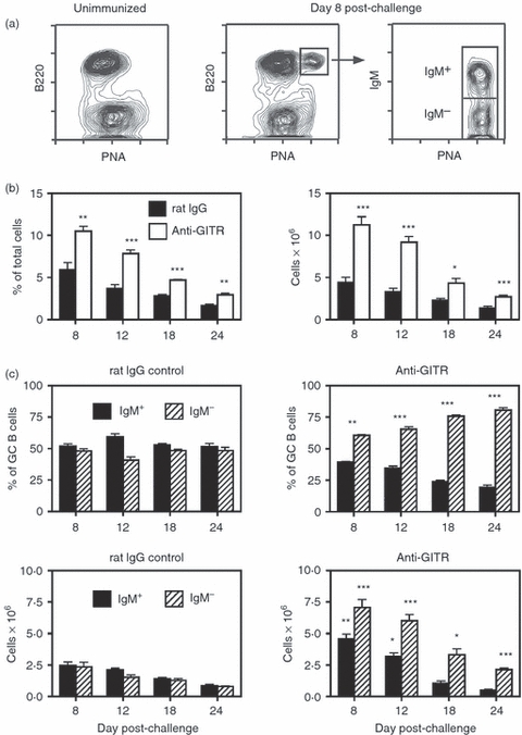
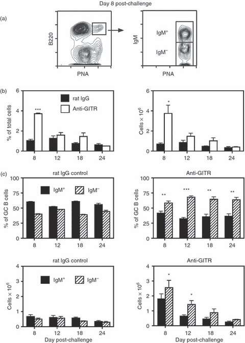
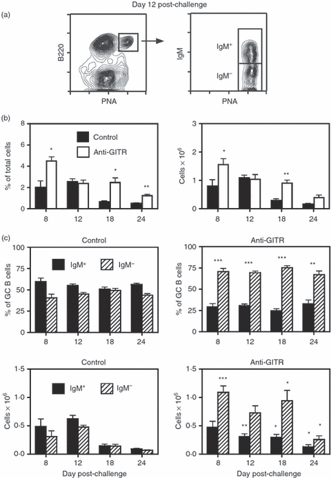
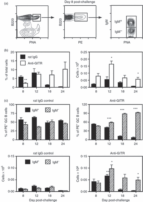
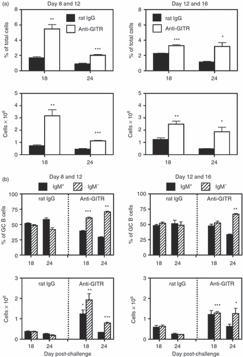
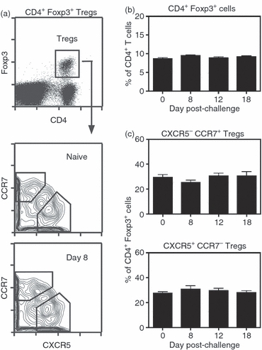
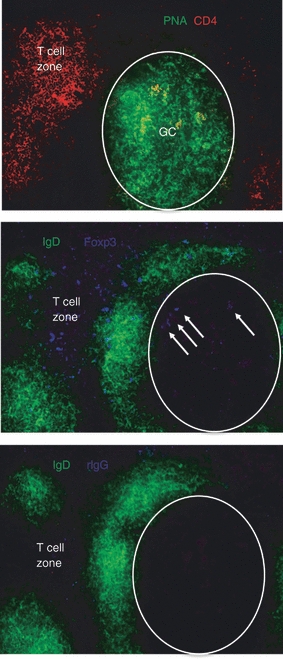
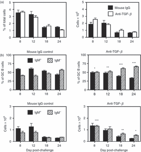
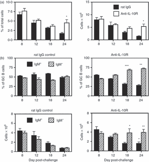
Germinal centre (GC) reactions are central features of T-cell-driven B-cell responses, and the site where antibody-producing cells and memory B cells are generated. Within GCs, a range of complex cellular and molecular events occur which are critical for the generation of high affinity antibodies. These processes require exquisite regulation not only to ensure the production of desired antibodies, but to minimize unwanted autoreactive or low affinity antibodies. To assess whether T regulatory (Treg) cells participate in the control of GC responses, immunized mice were treated with an anti-glucocorticoid-induced tumour necrosis factor receptor-related protein (GITR) monoclonal antibody (mAb) to disrupt Treg-cell activity. In anti-GITR-treated mice, the GC B-cell pool was significantly larger compared with control-treated animals, with switched GC B cells composing an abnormally high proportion of the response. Dysregulated GCs were also observed regardless of strain, T helper type 1 or 2 polarizing antigens, and were also seen after anti-CD25 mAb treatment. Within the spleens of immunized mice, CXCR5(+) and CCR7(-) Treg cells were documented by flow cytometry and Foxp3(+) cells were found within GCs using immunohistology. Final studies demonstrated administration of either anti-transforming growth factor-β or anti-interleukin-10 receptor blocking mAb to likewise result in dysregulated GCs, suggesting that generation of inducible Treg cells is important in controlling the GC response. Taken together, these findings indicate that Treg cells contribute to the overall size and quality of the humoral response by controlling homeostasis within GCs.


Figures

Figure 1
Figure 1
Disruption of regulatory T (Treg) cells leads to enhanced sheep red blood cell (SRBC)-induced germinal centre (GC) responses in BALB/c mice. Adult BALB/c mice were injected intraperitoneally (i.p.) with 250 μg of anti-glucocorticoid-induced tumour necrosis factor receptor-related protein (GITR) monoclonal antibody (mAb) or control rat IgG on days −2, +1 and +5. Mice were immunized on day 0 with SRBC, and spleens were harvested on days 8–24. (a) The left panel shows the absence of GC B cells [B220+ peanut agglutinin (PNA)hi] in unimmunized mice. The middle panel represents the splenic GC response in control rat IgG-treated mice 8 days post-challenge. The right panel illustrates IgM expression on the B220+ PNAhi GC population, with the IgM+ and IgM sub-sets designated. (b) The left panel shows the percent of B220+ PNAhi GC B cells within the viable lymphocyte gated population, and the right panel represents the number of total recovered splenic GC B cells at each time-point. Closed bars = control rat IgG-treated mice. Open bars = anti-GITR mAb-treated mice. Statistical analyses were performed between rat IgG and anti-GITR groups at each time-point. (c) The two upper panels show the percentages of IgM+ (solid bars) and IgM (hatched bars) B cells within the GC B-cell population. The two lower panels show the total number of IgM+ and IgM GC B cells within the spleen. Left panels are from control rat IgG-treated mice and right panels are from anti-GITR mAb-treated mice. Statistical analyses were performed between IgM+ GC B cells from rat IgG and anti-GITR groups and IgM GC B cells from rat IgG and anti-GITR groups at each time-point. Each bar represents average ± SD. There were six mice per group. *P < 0·04; **P < 0·005; ***P < 0·001.
Figure 2
Figure 2
Disruption of regulatory T (Treg) cells leads to enhanced sheep red blood cell (SRBC)-induced germinal centre (GC) responses in C57BL/6 mice. Adult C57BL/6 mice were injected intraperitoneally (i.p.) with 250 μg anti-glucocorticoid-induced tumour necrosis factor receptor-related protein (GITR) monoclonal antibody (mAb) or control rat IgG on days −2, +1 and +5. Mice were immunized on day 0 with SRBC, and spleens were harvested on days 8–24. (a) The left panel shows the splenic GC response in control rat IgG-treated mice 8 days post-challenge. The right panel illustrates IgM expression on the B220+ peanut agglutinin (PNA)hi GC population, with the IgM+ and IgM sub-sets designated. (b) The left panel shows the percentage of B220+ PNAhi GC B cells within the viable lymphocyte gated population, and the right panel represents the number of total recovered splenic GC B cells at each time-point. Closed bars = control rat IgG-treated mice. Open bars = anti-GITR mAb-treated mice. Statistical analyses were performed between rat IgG and anti-GITR groups at each time-point. (c) The two upper panels show the percentages of IgM+ (solid bars) and IgM (hatched bars) B cells within the GC B-cell population. The two lower panels show the total number of IgM+ and IgM GC B cells within the spleen. Left panels are from control rat IgG-treated mice and right panels are from anti-GITR mAb-treated mice. Statistical analyses were performed between IgM+ GC B cells from rat IgG and anti-GITR groups and IgM GC B cells from rat IgG and anti-GITR groups at each time-point. Each bar represents average ± SD. There were three to seven mice per group. *P < 0·04; **P < 0·02; ***P < 0·005.
Figure 3
Figure 3
Disruption of regulatory T (Treg) cells leads to enhanced influenza-induced germinal centre (GC) responses in BALB/c mice. Adult BALB/c mice were injected intraperitoneally (i.p.) with 250 μg anti-glucocorticoid-induced tumour necrosis factor receptor-related protein (GITR) monoclonal antibody (mAb) on days −2, +1 and +5. Mice were immunized on day 0 with influenza A virus (IAV; PR8, H1N1), and spleens were harvested on days 8–24. Mice immunized with IAV in the absence of any injections served as controls. (a) The left panel shows the splenic GC response in control-treated mice 12 days post-challenge. The right panel illustrates IgM expression on the B220+ peanut agglutinin (PNA)hi GC population, with the IgM+ and IgM sub-sets designated. (b) The left panel shows the percentage of B220+ PNAhi GC B cells within the viable lymphocyte gated population, and the right panel represents the number of total recovered splenic GC B cells at each time-point. Closed bars = control mice. Open bars = anti-GITR mAb-treated mice. Statistical analyses were performed between control and anti-GITR groups at each time-point. (c) The two upper panels show the percentages of IgM+ (solid bars) and IgM (hatched bars) B cells within the GC B-cell population. The two lower panels show the total number of IgM+ and IgM GC B cells within the spleen. Left panels are from control mice and right panels are from anti-GITR mAb-treated mice. Statistical analyses were performed between IgM+ GC B cells from control and anti-GITR groups and IgM GC B cells from control and anti-GITR groups at each time point. Each bar represents average ± SD. There were between five and 10 mice per group. *P < 0·04; **P < 0·004; ***P < 0·001.
Figure 4
Figure 4
Disruption of regulatory T (Treg) cells leads to enhanced phycoerythrin (PE)-induced germinal centre (GC) responses in BALB/c mice. Adult BALB/c mice were injected intraperitoneally (i.p.) with 250 μg anti-glucocorticoid-induced tumour necrosis factor receptor-related protein (GITR) monoclonal antibody (mAb) or control rat IgG on days −2, +1 and +5. Mice were immunized on day 0 with phycoerythrin (PE) precipitated in alum, and spleens were harvested on days 8–24. Given the relatively low frequency of PE-binding GC B cells, at least 2–3 × 10⁶ events were collected per sample during flow cytometric analysis. (a) The left panel shows the splenic GC response in anti-GITR mAb-treated mice 8 days post-challenge. The middle panel illustrates the PE-binding B cells within the B220+ peanut agglutinin (PNA)hi GC population. The right panel shows the IgM+ and IgM sub-sets within the PE-binding GC B cells. (b) The left panel shows the percent of PE-binding B220+ PNAhi GC B cells within the viable lymphocyte gated population, and the right panel represents the number of total recovered splenic PE-binding GC B cells at each time point. Closed bars = control rat IgG treated mice. Open bars = anti-GITR mAb treated mice. Statistical analyses were performed between rat IgG and anti-GITR groups at each time point. (c) The two upper panels show the percentages of IgM+ (solid bars) and IgM (hatched bars) B cells within the PE-binding B220+ PNAhi GC B-cell population. The two lower panels show the total number of IgM+ and IgM PE-binding GC B cells within the spleen. Left panels are from control rat IgG-treated mice and right panels are from anti-GITR mAb-treated mice. Statistical analyses were performed between IgM+ PE-binding GC B cells from rat IgG and anti-GITR groups and IgM PE-binding GC B cells from rat IgG and anti-GITR groups at each time-point. Each bar represents average ± SD. There were four to 11 mice per group. *P < 0·02; **P < 0·002; ***P < 0·0001.
Figure 5
Figure 5
T regulatory (Treg) cells influence the germinal centre (GC) response at late time-points. BALB/c mice were immunized with sheep red blood cells (SRBC) on day 0, and injected intraperitoneally (i.p.) with 250 μg of anti-glucocorticoid-induced tumour necrosis factor receptor-related protein (GITR) monoclonal antibody (mAb) or control rat IgG at either days 8 and 12 or days 12 and 16 post-challenge, and spleens were harvested on days 18 and 24. (a) Left panels are from mice injected on days 8 and 12 with anti-GITR mAb or control rat IgG, and right panels are mice injected on days 12 and 16. Upper panels show the percentage of B220+ peanut agglutinin (PNA)hi GC B cells within the viable lymphocyte gated population, and the lower panels represent the number of total recovered splenic GC B cells at each time-point. Closed bars = control rat IgG-treated mice. Open bars = anti-GITR mAb-treated mice. Statistical analyses were performed between rat IgG and anti-GITR groups at each time point. (b) Left panels are from mice injected on days 8 and 12 with anti-GITR mAb or control rat IgG, and right panels are mice injected on days 12 and 16. The two upper panels show the percentages of IgM+ (solid bars) and IgM (hatched bars) B cells within the GC B-cell population. The two lower panels show the total number of IgM+ and IgM GC B cells within the spleen. Statistical analyses were performed between IgM+ GC B cells from rat IgG and anti-GITR groups and IgM GC B cells from rat IgG and anti-GITR groups at each time point. Each bar represents average ± SD. There were three or four mice per group. *P < 0·04; **P < 0·02; ***P < 0·005.
Figure 6
Figure 6
Chemokine receptor expression on splenic CD4+ Foxp3+ T cells. B6.FoxP3-GFP mice were immunized with sheep red blood cells (SRBC) on day 0 and spleens were harvested on days 8, 12 and 18. Cells were stained with anti-CD4, anti-CXCR5 and anti-CCR7 monoclonal antibodies (mAbs). (a) The upper panel shows the gated CD4+ GFP (Foxp3)+ regulatory T (Treg) cell sub-set. The lower panels show Treg-cell sub-sets defined by CXCR5 and CCR7 in naive (day 0) and day 8 challenged mice. The gated CXCR5 CCR7+ and CXCR5+ CCR7 sub-sets are indicated. (b) Percentage of CD4+ Foxp3+ cells within the splenic CD4+ T-cell compartment of either naive or immunized mice. (c) Percentage of CXCR5 CCR7+ and CXCR5+ CCR7 cells within the splenic CD4+ Foxp3+ Treg-cell population of naive and immunized mice. There were three to six mice per group.
Figure 7
Figure 7
Foxp3+ cells are physically present within splenic germinal centres (GCs). Adult BALB/c mice were injected intraperitoneally (i.p.) with sheep red blood cells (SRBC) on day 0, and spleens were harvested on day 8. Splenic fragments were snap frozen, thin sectioned (8 μm) and serial sections were stained with anti-CD4 monoclonal antibody (mAb) and peanut agglutinin (PNA), or anti-IgD mAb and either anti-Foxp3 mAb or rat IgG2a isotype control. Sections were imaged using a Nikon Eclipse E600 fluorescence microscope equipped with a SPOT digital camera, and images were processed with Adobe Photoshop software. The upper panel shows CD4 (red/Cy3) and PNA (green/FITC) staining. The lower panels show IgD (green/FITC) and Foxp3 or isotype control (blue/Cy5) staining. The areas composing GCs are outlined in the lower panels. Foxp3+ cells within the GC are indicated by arrows in the middle panel. Magnification 100 ×. Images are representative of multiple fields from three mice. Please note that the Foxp3 and isotype control files were imaged identically with the SPOT digital camera, and were similarly colour enhanced identically in Photoshop.
Figure 8
Figure 8
Neutralization of transforming growth factor-β (TGF-β) leads to enhanced sheep red blood cell (SRBC) -induced germinal centre (GC) responses. Adult BALB/c mice were injected intraperitoneally (i.p.) with 100 μg of anti-TGF-β (1D11) monoclonal antibody (mAb) or control mouse IgG every 2 days starting at day 0 and continued until the mice were killed. Mice were immunized on day 0 with SRBC, and spleens were harvested on days 8–24. (a) The left panel shows the percentage of B220+ peanut agglutinin (PNA)hi GC B cells within the viable lymphocyte gated population, and the right panel represents the number of total recovered splenic GC B cells at each time-point. Closed bars = control mouse IgG-treated mice. Open bars = anti-TGF-β mAb treated mice. Statistical analyses were performed between mouse IgG and anti-TGF-β groups at each time-point. (b) The two upper panels show the percentages of IgM+ (solid bars) and IgM (hatched bars) B cells within the GC B-cell population. The two lower panels show the total number of IgM+ and IgM GC B cells within the spleen. Left panels are from control mouse IgG-treated mice and right panels are from anti-TGF-β mAb-treated mice. Statistical analyses were performed between IgM+ GC B cells from mouse IgG and anti-TGF-β groups and IgM GC B cells from mouse IgG and anti-TGF-β groups at each time-point. Each bar represents average ± SD. There were five or six mice per group. *P < 0·03; **P < 0·015; ***P < 0·01.
Figure 9
Figure 9
Blockade of interleukin-10 receptor (IL-10R) leads to enhanced sheep red blood cell (SRBC)-induced germinal centre (GC) responses. Adult BALB/c mice were injected intraperitoneally (i.p.) with 1 mg of anti-IL-10R (1B1.3a) mAb and control rat IgG on day 0, and twice weekly with 500 μg until the mice were killed, starting in the second week. Mice were immunized on day 0 with SRBC, and spleens were harvested on days 8–24. (a) The left panel shows the percentage of B220+ peanut agglutinin (PNA)hi GC B cells within the viable lymphocyte-gated population, and the right panel represents the number of total recovered splenic GC B cells at each time-point. Closed bars = control rat IgG-treated mice. Open bars = anti-IL-10R mAb-treated mice. Statistical analyses were performed between rat IgG and anti-IL-10R groups at each time-point. (b) The two upper panels show the percentages of IgM+ (solid bars) and IgM (hatched bars) B cells within the GC B-cell population. The two lower panels show the total number of IgM+ and IgM GC B cells within the spleen. Left panels are from control rat IgG-treated mice and right panels are from anti-IL-10R mAb-treated mice. Statistical analyses were performed between IgM+ GC B cells from rat IgG and anti-IL-10R groups and IgM GC B cells from rat IgG and anti-IL-10R groups at each time-point. Each bar represents average ± SD. There were thre to six mice per group. *P < 0·05; **P < 0·03; ***P < 0·005.

References

    1. Wolniak KL, Shinall SM, Waldschmidt TJ. The germinal center response. Crit Rev Immunol. 2004;24:39–65. - PubMed
    1. Allen CD, Okada T, Cyster JG. Germinal-center organization and cellular dynamics. Immunity. 2007;27:190–202. - PMC - PubMed
    1. Klein U, Dalla-Favera R. Germinal centres: role in B-cell physiology and malignancy. Nat Rev Immunol. 2008;8:22–33. - PubMed
    1. Goodnow CC, Vinuesa CG, Randall KL, Mackay F, Brink R. Control systems and decision making for antibody production. Nat Immunol. 2010;11:681–8. - PubMed
    1. Shinall SM, Gonzalez-Fernandez M, Noelle RJ, Waldschmidt TJ. Identification of murine germinal center B cell subsets defined by the expression of surface isotypes and differentiation antigens. J Immunol. 2000;164:5729–38. - PubMed

Publication types

LinkOut - more resources