Comparative Study
. 2011 Jun 7;57(23):2317-27.
doi: 10.1016/j.jacc.2010.12.036.

Distinguishing arrhythmogenic right ventricular cardiomyopathy/dysplasia-associated mutations from background genetic noise

Affiliations
Comparative Study

Distinguishing arrhythmogenic right ventricular cardiomyopathy/dysplasia-associated mutations from background genetic noise

Jamie D Kapplinger et al. J Am Coll Cardiol. .

Abstract

Objectives: The aims of this study were to determine the spectrum and prevalence of "background genetic noise" in the arrhythmogenic right ventricular cardiomyopathy/dysplasia (ARVC) genetic test and to determine genetic associations that can guide the interpretation of a positive test result.

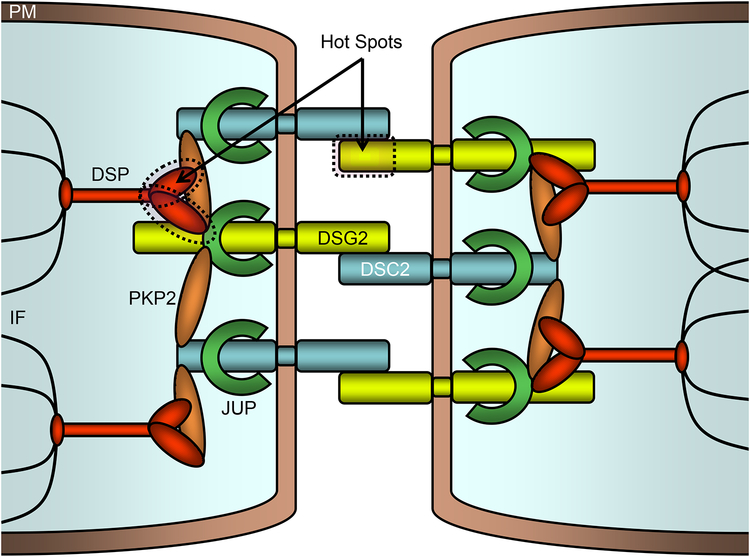
Background: ARVC is a potentially lethal genetic cardiovascular disorder characterized by myocyte loss and fibrofatty tissue replacement of the right ventricle. Genetic variation among the ARVC susceptibility genes has not been systematically examined, and little is known about the background noise associated with the ARVC genetic test.

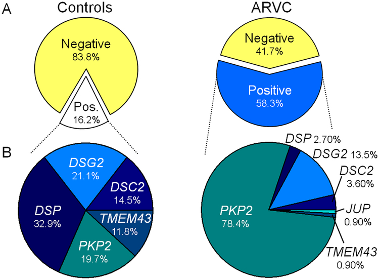
Methods: Using direct deoxyribonucleic acid sequencing, the coding exons/splice junctions of PKP2, DSP, DSG2, DSC2, and TMEM43 were genotyped for 93 probands diagnosed with ARVC from the Netherlands and 427 ostensibly healthy controls of various ethnicities. Eighty-two additional ARVC cases were obtained from published reports, and additional mutations were included from the ARVD/C Genetic Variants Database.

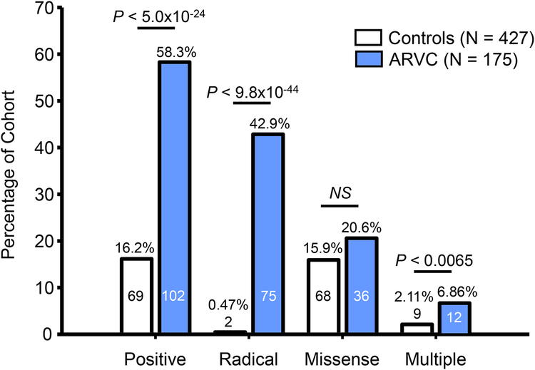
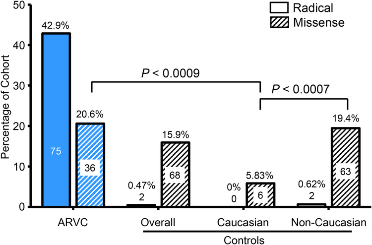
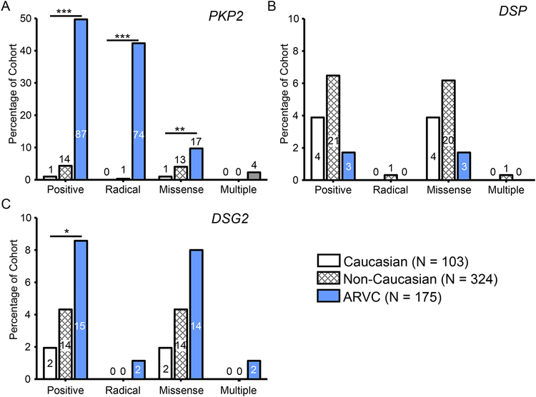
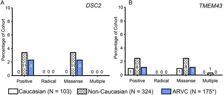
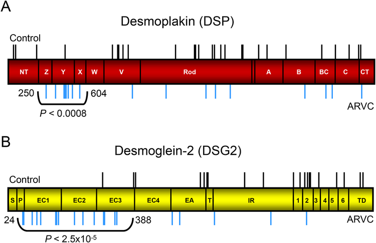
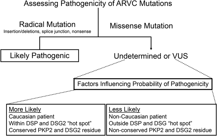
Results: The overall yield of mutations among ARVC cases was 58% versus 16% in controls. Radical mutations were hosted by 0.5% of control individuals versus 43% of ARVC cases, while 16% of controls hosted missense mutations versus a similar 21% of ARVC cases. Relative to controls, mutations in cases occurred more frequently in non-Caucasians, localized to the N-terminal regions of DSP and DSG2, and localized to highly conserved residues within PKP2 and DSG2.

Conclusions: This study is the first to comprehensively evaluate genetic variation in healthy controls for the ARVC susceptibility genes. Radical mutations are high-probability ARVC-associated mutations, whereas rare missense mutations should be interpreted in the context of race and ethnicity, mutation location, and sequence conservation.


Figures

Figure 1 –
Figure 1 –. Prevalence of mutations in control and ARVC cohorts.
A) Pie charts of the percentage of mutation-positive individuals within the control (white) and ARVC cohorts (blue) separated from mutation-negative individuals (yellow) of each respective cohort. B) Pie chart of the percentage of mutations found in each of the ARVC-associated genes among mutation-positive cases in the control (left) and ARVC (right) cohort. JUP was not genotyped in control cases and TMEM43 was not genotyped in the United States ARVD/C Registry component of the ARVC case cohort.
Figure 2 –
Figure 2 –. Prevalence of radical, missense, and multiple mutations.
Bar graph depicting the number and percentage of the control (white) and ARVC case (blue) cohorts hosting mutations. The overall yield of the genetic test (positive) was divided into radical mutations, missense mutations, and individuals hosting more than one mutation (multiple). Numbers within bars denote number of individuals positive for the respective mutation type in each cohort.
Figure 3 –
Figure 3 –. Prevalence of mutations in Caucasian and non-Caucasian individuals.
Bar graph of the number and percentage of the ARVC (blue background) or control (white background) individuals hosting radical (no lines) or missense (diagonal lines) mutations. Mutations identified in controls were subdivided into Caucasian and non-Caucasian ostensibly healthy individuals. Numbers within bars denote number of individuals positive for the respective mutation type in each cohort and cohort subset.
Figure 4 –
Figure 4 –. Prevalence of mutations in PKP2, DSP, and DSG2.
Bar graph depicting the number and percentage of the Caucasian control (white), non-Caucasian control (white with hash) and ARVC case (blue) cohorts hosting mutations in PKP2 (A), DSP (B), and DSG2 (C). The overall yield of the genetic test (positive) was divided into radical mutations, missense mutations, and individuals hosting more than one mutation (multiple). Numbers within bars denote number of individuals positive for a mutation in the respective gene in each cohort. *, P < 0.05; **, P < 0.01; ***, P < 0.0001.
Figure 5 –
Figure 5 –. Prevalence of mutations in DSC2 and TMEM43.
Bar graph depicting the number and percentage of the Caucasian control (white), non-Caucasian control (white with hash) and ARVC case (blue) cohorts hosting mutations in DSC2 (A) and TMEM43 (B). The overall yield of the genetic test (positive) was divided into radical mutations, missense mutations, and individuals hosting more than one mutation (multiple). Numbers within bars denote number of individuals positive for a mutation in the respective gene in each cohort. *, TMEM43 was not genotyped in the United States ARVD/C Registry component of the ARVC case cohort (N = 93).
Figure 6 –
Figure 6 –. DSP and DSG mutation “hot spots”.
Linear topology of DSP and DSG2 with the location of all control missense mutations (black line, above topology) and ARVC case mutations (blur lines, below topology) identified. A) ARVC mutations were more frequent between residues 250 and 604 of DSP (red topology) than in control individuals. NT, Z, Y, X, W, V, predicted alpha-helical bundles; A, B, BC, C, CT, intermediate filament binding domains. B) ARVC mutations were more frequent between residues 24 and 388 of DSG2 (yellow topology) than in control individuals. S, signal peptide; P, propeptide; EC1, 2, 3, 4, cadherin repeat domains I-IV; EA, extracellular anchor; TM, transmembrane domain; IA, intracellular anchor; ICS, intracellular catenin-binding domain; IPL, proline-rich linker; RUD, repeating unit domain; DTD, desmoglein terminal domain.
Figure 7 –
Figure 7 –. Mutation “hot spots” within the cardiocyte desmosome.
Schematic representation of the desmosome which links neighboring cellular intermediate filaments through a complex of interacting proteins. Two mutation hotspots (dashed outline) on DSP and DSG2 are noted. PM, plasma membrane; DSP, desmoplakin; DSG2, desmoglein 2; DSC2, desmocolin 2; PKP2, plakophilin; JUP, plakoglobin 2; IF, intermediate filaments.
Figure 8 –
Figure 8 –. Assessing likelihood of pathogenicity of a positive ARVC genetic test.
Flow chart of factors influencing likelihood that a given ARVC genetic test-identified mutation is pathogenic based on comparison of mutations identified in ostensibly healthy controls versus definite ARVC cases.

Comment in

References

    1. Peters S, Trümmel M, Meyners W. Prevalence of right ventricular dysplasia-cardiomyopathy in a non-referral hospital. Int. J. Cardiol 2004; 97:499–501. - PubMed
    1. Gemayel C, Pelliccia A, Thompson PD. Arrhythmogenic right ventricular cardiomyopathy. J Am Coll Cardiol. 2001; 38:1773–1781. - PubMed
    1. Dalal D, Molin LH, Piccini J, Tichnell C, James C, Bomma C, Prakasa K, Towbin JA, Marcus FI, Spevak PJ, Bluemke DA, Abraham T, Russell SD, Calkins H, Judge DP. Clinical Features of Arrhythmogenic Right Ventricular Dysplasia/Cardiomyopathy Associated With Mutations in Plakophilin-2. Circulation. 2006; 113:1641–1649. - PubMed
    1. Sen-Chowdhry S, Syrris P, McKenna WJ. Role of Genetic Analysis in the Management of Patients With Arrhythmogenic Right Ventricular Dysplasia/Cardiomyopathy. J Am Coll Cardiol. 2007; 50:1813–1821. - PubMed
    1. Thiene G, Corrado D, Basso C. Arrhythmogenic right ventricular cardiomyopathy/dysplasia. Orphanet J Rare Dis. 2007; 2:45. - PMC - PubMed

Publication types