. 2011 Sep;32(26):6068-79.
doi: 10.1016/j.biomaterials.2011.05.006. Epub 2011 Jun 1.

Robust CNS regeneration after complete spinal cord transection using aligned poly-L-lactic acid microfibers

Affiliations

Robust CNS regeneration after complete spinal cord transection using aligned poly-L-lactic acid microfibers

Andres Hurtado et al. Biomaterials. 2011 Sep.

Abstract

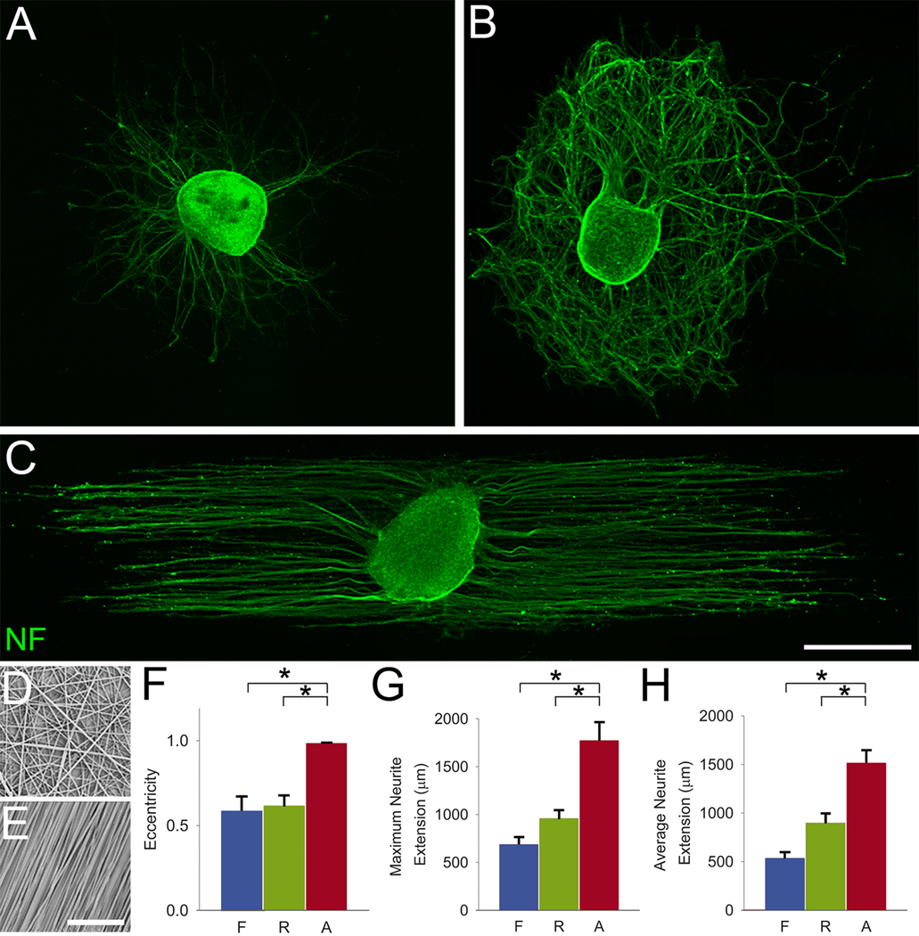
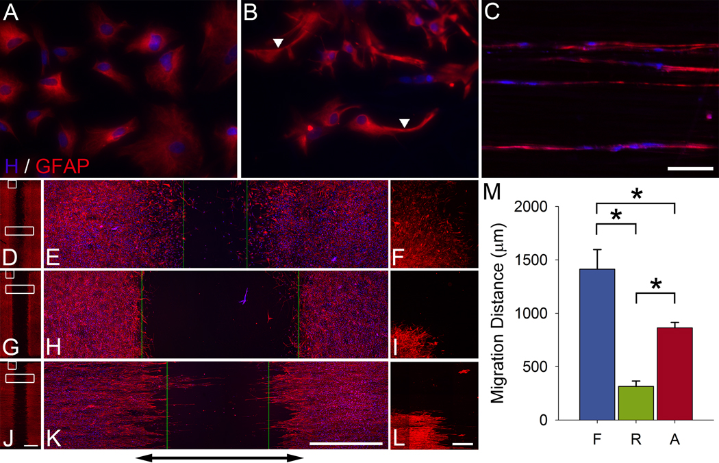
Following spinal cord injury, axons fail to regenerate without exogenous intervention. In this study we report that aligned microfiber-based grafts foster robust regeneration of vascularized CNS tissue. Film, random, and aligned microfiber-based conduits were grafted into a 3 mm thoracic rat spinal cord gap created by complete transection. Over the course of 4 weeks, microtopography presented by aligned or random poly-L-lactic acid microfibers facilitated infiltration of host tissue, and the initial 3 mm gap was closed by endogenous cell populations. This bulk tissue response was composed of regenerating axons accompanied by morphologically aligned astrocytes. Aligned fibers promoted long distance (2055 ± 150 μm), rostrocaudal axonal regeneration, significantly greater than random fiber (1162 ± 87 μm) and film (413 ± 199 μm) controls. Retrograde tracing indicated that regenerating axons originated from propriospinal neurons of the rostral spinal cord, and supraspinal neurons of the reticular formation, red nucleus, raphe and vestibular nuclei. Our findings outline a form of regeneration within the central nervous system that holds important implications for regeneration biology.


Figures

Fig. 1
Fig. 1
Schematic detailing materials fabrication process and characterization. A custom electrospinning apparatus (A) was used to generate aligned polymeric fibers. Coverslips were mounted on a grounded target, and a rotation speed of 1500 rpm was used to align fibers produced by a 15 kV field potential (B). Random fibers were generated using a stationary target. For conduit assembly, films with or without electrospun fibers were peeled from coverslips (C), placed back to back (D), and rolled (E) into conduits (F). Random (G) and aligned (I) fibers were visualized by scanning electron microscopy, and alignment was quantified by measuring the angle between a given fiber and the median fiber orientation for 150 fibers per condition (H and J, respectively). Importantly, fiber alignment was maintained through the process of conduit assembly (K and L). (M) Macroscopic view of aligned fiber conduit lumen, visualized by mounting a conduit sectioned on the longitudinal axis. (N) Coronal view of an aligned fiber conduit, the diameter of all conduits was 2.6 mm. Scale bars: 50 µm in (G); 100 µm in (I and K); 1 mm in (M and N).
Fig. 2
Fig. 2
Aligned polymer fibers specify the direction of DRG neurite growth. DRG isolated from P4 rat pups were cultured on film (A), random (B), and aligned fiber (C) substrates. Random (D) and aligned (E) polymer fibers were characterized by scanning electron microscopy. The eccentricity (F), a measure of anisotropy, and the maximum (G) and average (H) distance reached by neurites were quantified on DRG explants. Markedly, aligned polymer fibers elicited linear neurite elongation, providing an efficient means of growth as demonstrated by a significant increase in the maximum and average distance reached by neurites over the same time in culture. F, film; R, random; A, aligned. Data are mean ± SEM; n = 6. *P < 0.05 by ANOVA. Scale bars: A–C, 500 µm; D, E, 50 µm.
Fig. 3
Fig. 3
Microtopography promotes host tissue integration and gap closure. A 3 mm long conduit was implanted to bridge a complete transection spinal cord injury. Cresyl violet was used to visualize tissue architecture. Representative images 4 weeks after implantation of film (A), random (B), and aligned fiber (C) conduits demonstrate that both random and aligned fibers support tissue integration into conduits and limited cavitation in host cord tissue. A Cavalieri estimator probe was used to measure tissue volume inside conduits at 1, 2, and 4 weeks post-injury (D). Both random and aligned fiber conduits had significantly more endogenous tissue than film conduits at 4 weeks. Data are mean ± SEM; n = 3 (1W, 2W), n = 7 (4W). *P < 0.05 by ANOVA. Scale bar: A–C, 1 mm.
Fig. 4
Fig. 4
Grafts support angiogenesis and are well vascularized 4 weeks following conduit implantation. Blood vessels are observed, by immunostaining for rat endothelial cell antigen 1 (RECA-1), at the rostral cord interface 1 week after implantation (A), and in the center of the graft at 4 weeks (B). C, Immunostaining for laminin in the basement membrane of blood vessels. A distinct difference in the pattern of vascularization is observed between the rostral (D) and caudal (E) spinal cord. In the rostral spinal cord, blood vessel formation occurs in close proximity to the regeneration front. D, E, Insets from (C). Scale bars: A, 100 µm; B, 50 µm; C, 500 µm; D, E, 200 µm.
Fig. 5
Fig. 5
Aligned fibers promote extensive axonal regeneration. Immunostaining for neurofilament (RT97) was used to visualize axons. Representative horizontal spinal cord sections for film (A, D, G), random (B, E, H), and aligned fiber (C, F, I) conduits. Aligned fibers foster robust, time-dependent rostrocaudal axonal regeneration (C, F, I), whereas the same response is absent in film and random fiber conduits. Dotted lines indicate the walls of the conduits. Arrowheads (E, F, H, I) indicate the regeneration front. J, The axonal regeneration response inside aligned conduits was markedly linear, shown here in a different animal than that presented in (I). Serotonergic (5HT+) axons were abundant in the robust growth observed inside aligned conduits (K, inset from adjacent section of the same animal in I). L, Serotonergic axons were present caudal to the graft in 3/21 animals (2 random, 1 aligned fiber). The distance between the rostral edge of the conduit to the ‘axonal front’ was quantified at all time points (M). Remarkably, over 4 weeks, aligned fibers promote robust, long distance regeneration (2055 ± 150 µm), significantly greater than random fiber (1162 ± 87 µm) and film (413 ± 199 µm) controls. Notably, at 4 weeks, 100% (7/7) of the animals from the aligned fiber group had a robust regeneration response present in the middle of the conduit compared to 14.3% (1/7) and 0% (0/6) in the random fiber and film groups, respectively (N). Data are mean ± SEM; n = 3 (1W, 2W), n = 6–7 (4W). *P < 0.05 by ANOVA. Scale bars: A–I, 1 mm; J, 500 µm; K, 150 µm; L, 50 µm.
Fig. 6
Fig. 6
Axonal regeneration is localized to migratory astrocytes. GFAP staining was used to visualize astrocytes. Representative horizontal spinal cord sections for film (A, D, G), random (B, E, H), and aligned fiber (C, F, I) conduits. Both random and aligned fibers support a time-dependent migratory response of astrocytes (B, C, E, F, H, I), whereas time-dependent astrocytic dieback is observed in film conduits (D, G). Double-labeling for GFAP and RT97 indicates axonal regeneration is localized to astrocytes migrating from the rostral spinal cord (J, inset from I). High-magnification confocal micrographs show morphologically aligned migrating astrocytes in close proximity to regenerating axons (K and L, inset from J). Scale bars: A–I, 1 mm; J, 500 µm; K, L, 50 µm.
Fig. 7
Fig. 7
Astrocytes exhibit morphological sensitivity to topographic cues and migrate in a topography dependent manner. Astrocytes isolated from neonatal rat cortices were cultured for 48 h on film (A), random (B), and aligned fiber (C) substrates. Arrows indicate close apposition of astrocytes to underlying polymer fibers in (B). Astrocytes exhibited a linear morphology when cultured on aligned fibers (C). In a migration assay (D–L), astrocytes cultured on film and aligned fibers partially close the initial 2.25 mm gap (indicated by black arrow), whereas astrocytes cultured on random fibers remain relatively static over 5 days in culture (E, F, insets from D; H, I, insets from G; K, L, insets from J). Green lines in E, H, and K indicate the migration fronts. Interestingly, astrocytes are only able to migrate on aligned fibers in a direction parallel to the median fiber orientation (L). Migration of astrocytes on film is significantly greater than migration of astrocytes on random and aligned fibers, and migration of astrocytes on aligned fibers is significantly greater than migration of astrocytes on random fibers (M). Data are mean ± SEM; n = 10. *P < 0.05 by ANOVA. Scale bars: A–C, 100 µm; D, G, J, 2 mm; E, F, H, I, K, L, 1 mm.
Fig. 8
Fig. 8
Pioneer axons guide the regeneration response from the rostral spinal cord. A, D, Cresyl violet staining in sections adjacent to GFAP / RT97 stained sections demonstrate that polymer fibers guide rostrocaudal growth in random (A) and aligned fiber (D) conduits. Black arrowheads point to polymer fibers, random fibers in (A) project orthogonal to the plane of the tissue section, whereas aligned fibers (D) remain in the plane of the section. B, E, After 4 weeks, astrocytes (GFAP) and regenerating axons (RT97) enter random (B) and aligned fiber (E) conduits in close proximity, and axons lead the response. Dotted lines indicate the walls of the conduits. White arrowheads point to axons at the regeneration front. C, Inset from (B). F, Inset from (E). Scale bars: B, E, 500 µm; E, F, 200 µm.
Fig. 9
Fig. 9
Axon growth precedes astrocyte migration from cortical explants in vitro. Cortical explants were isolated from E18 rat pups and cultured on film (A), random (B), and aligned (C) fiber substrates for 5 days. D, E, F, Insets of astrocyte migration fronts from cortical explants (D, inset from A; E, inset from B; F, inset from C) demonstrate that axonal growth from cortical explants is ahead of astrocyte migration. Scale bars: A–C, 500 µm; D–F, 250 µm.

References

    1. Silver J, Miller JH. Regeneration beyond the glial scar. Nat Rev Neurosci. 2004;5:146–156. - PubMed
    1. Ylera B, Ertürk A, Hellal F, Nadrigny F, Hurtado A, Tahirovic S, et al. Chronically CNS-injured adult sensory neurons gain regenerative competence upon a lesion of their peripheral axon. Curr Biol. 2009;19:930–936. - PubMed
    1. Hellal F, Hurtado A, Ruschel J, Flynn KC, Laskowski CJ, Umlauf M, et al. Microtubule stabilization reduces scarring and causes axon regeneration after spinal cord injury. Science. 2011;331:928–931. - PMC - PubMed
    1. Moore DL, Blackmore MG, Hu Y, Kaestner KH, Bixby JL, Lemmon VP, et al. KLF family members regulate intrinsic axon regeneration ability. Science. 2009;326:298–301. - PMC - PubMed
    1. Neumann S, Bradke F, Tessier-Lavigne M, Basbaum AI. Regeneration of sensory axons within the injured spinal cord induced by intraganglionic cAMP elevation. Neuron. 2002;34:885–893. - PubMed

Publication types