. 2011 Jun 1;17(11):3590-9.
doi: 10.1158/1078-0432.CCR-10-3349.

BRAF activation induces transformation and then senescence in human neural stem cells: a pilocytic astrocytoma model

Affiliations

BRAF activation induces transformation and then senescence in human neural stem cells: a pilocytic astrocytoma model

Eric H Raabe et al. Clin Cancer Res. .

Abstract

Purpose: BRAF is frequently activated by gene fusion or point mutation in pilocytic astrocytoma, the most common pediatric brain tumor. We investigated the functional effect of constitutive BRAF activation in normal human neural stem and progenitor cells to determine its role in tumor induction in the brain.

Experimental design: The constitutively active BRAF(V600E) allele was introduced into human neurospheres, and its effects on MAPK (mitogen-activated protein kinase) signaling, proliferation, soft agarose colony formation, stem cell phenotype, and induction of cellular senescence were assayed. Immunohistochemistry was used to examine p16(INK4a) levels in pilocytic astrocytoma.

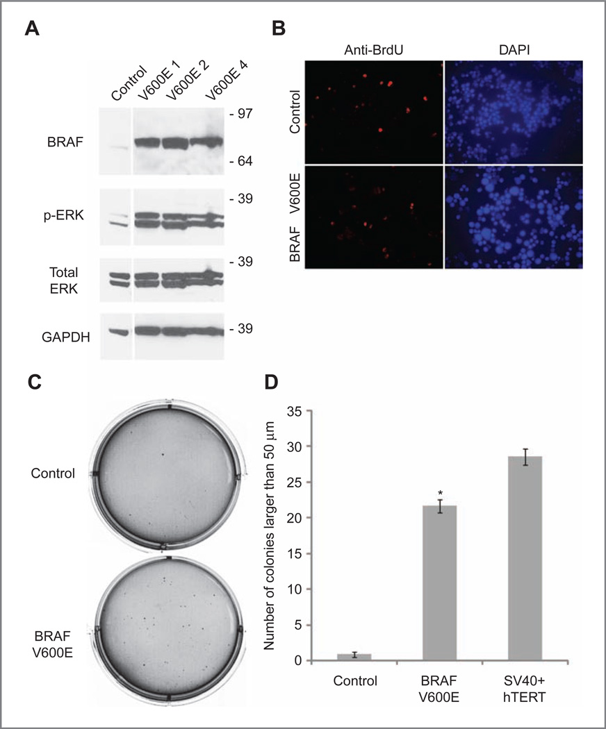
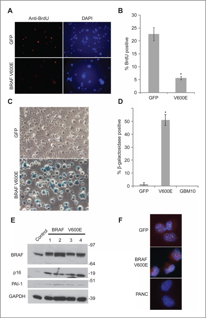
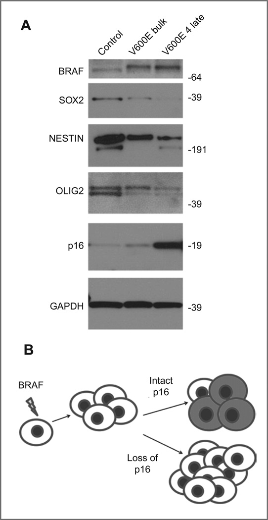
Results: BRAF(V600E) expression initially strongly promoted colony formation but did not lead to significantly increased proliferation. BRAF(V600E)-expressing cells subsequently stopped proliferating and induced markers of oncogene-induced senescence including acidic β-galactosidase, PAI-1, and p16(INK4a) whereas controls did not. Onset of senescence was associated with decreased expression of neural stem cell markers including SOX2. Primary pilocytic astrocytoma cultures also showed induction of acidic β-galactosidase activity. Immunohistochemical examination of 66 pilocytic astrocytomas revealed p16(INK4a) immunoreactivity in the majority of cases, but patients with tumors negative for p16(INK4a) had significantly shorter overall survival.

Conclusions: BRAF activation in human neural stem and progenitor cells initially promotes clonogenic growth in soft agarose, suggesting partial cellular transformation, but oncogene-induced senescence subsequently limits proliferation. Induction of senescence by BRAF may help explain the low-grade pathobiology of pilocytic astrocytoma, whereas worse clinical outcomes associated with tumors lacking p16(INK4a) expression could reflect failure to induce senescence or an escape from oncogene-induced senescence.


Conflict of interest statement

Disclosure of Potential Conflicts of Interest

No potential conflicts of interest were disclosed.

Figures

Figure 1
Figure 1
ERK activation, cellular proliferation, and colony formation in soft agarose following BRAFV600E transduction. A, Western blot showing that normal human neural stem and progenitor cells transduced with BRAFV600E lentivirus express higher levels of activated p-ERK than control neurospheres. V600E 1–4 represent separate subclones transduced with BRAFV600E. B, proliferation as measured by BrdU incorporation is initially similar between control cells (top) and BRAFV600E-infected cells (bottom), with 11% of control cells BrdU positive and 13% of BRAFV600E cells BrdU positive, P = 0.33, Student's t test. C, soft agarose experiment, showing that control cells (top) do not form large numbers of colonies compared with BRAFV600E-transduced cells. D, quantification of soft agarose colony formation, including SV40 large T antigen and hTERT-transduced cells as positive controls. BRAFV600E-infected cells show statistically significant increase in colony formation compared with control cortex neurosphere cells. *, P < 0.0001, control versus BRAFV600E-infected cells, Student's t test.
Figure 2
Figure 2
BRAFV600E-expressing cells upregulate senescence markers and show decreased proliferation after several passages. A, immunofluorescence 200× photomicrographs of BrdU (red) and DAPI (blue)-stained GFP control and BRAFV600E-expressing neural stem and progenitor cells. After 5 passages, BRAFV600E-expressing neurosphere cells show increased nuclear size (DAPI) and decreased proliferation as measured by BrdU incorporation (bottom) compared with control neurospheres (top). B, quantification of BrdU positivity, showing percentage of BrdU-positive cells. *, P < 0.05, GFP versus BRAFV600E-infected cells, Student's t test. C, bright field 200× photomicrographs of acidic β-galactosidase activity in control GFP and BRAFV600E-infected neurosphere cells. Cells have been plated on an adherent substrate to allow for improved visualization of acidic β-galactosidase staining. Blue staining, indicating increased acidic β-galactosidase activity, is increased in BRAFV600E-transduced cells, which also become enlarged when compared with GFP-expressing controls. D, quantification of acidic β-galactosidase activity, comparing the mean percentage of β-galactosidase–positive cells in GFP control and BRAFV600E-transduced neural stem and progenitor cells. *, P < 0.005 GFP versus BRAFV600E-infected cells, Student's t test. E, Western blotting showing increased p16INK4a and PAI-1 expression in late-passage BRAFV600E-expressing cells compared with controls. Senescent markers p16INK4a and PAI-1 are induced in late-passage BRAFV600E-expressing neurosphere cells compared with control neurosphere cells. BRAFV600E 1–4 represent subclones isolated from the bulk infected culture. Molecular weight markers are located at the right of the blot. F, FISH analysis reveals similar telomere length in GFP control and BRAFV600E-expressing neurosphere cells. Fluorescent 1000× photomicrographs of FISH-stained nuclei from similar passage GFP and BRAFV600E neurospheres. At this passage, BRAFV600E-transduced cells were highly senescent whereas GFP-expressing neurospheres continued to proliferate normally. Telomeres are labeled red, and centromeres are labeled green. Nuclei are counterstained with DAPI. PANC is a pancreatic carcinoma cell line with shortened telomeres and corresponding decreased red telomere signal.
Figure 3
Figure 3
Pilocytic astrocytoma cells express markers of senescence in vivo and in vitro. Photomicrographs showing p16INK4a immunohistochemical staining in pilocytic astrocytoma specimens: negative (A); weak (B); moderate (C); and strong (D) p16INK4a immunohistochemical staining in pilocytic astrocytomas. E and F, immunohistochemical staining for p53 was largely negative, with only rare tumors showing scattered weakly staining cells. F, digitally expanded higher power view showing nuclear p53 staining in elongated tumor cell. (Original magnification 400× for A–F.) G, log-rank analysis of Kaplan–Meier curves showing significantly shorter overall survival in patients whose tumors were p16INK4a immunonegative (P = 0.0002). H and I, acidic β-galactosidase staining of an early-passage representative primary pilocytic astrocytoma culture. Bright field photomicrograph (100× power) showing prominent acidic β-galactosidase activity in primary pilocytic astrocytoma cells after 3 passages in culture (I), whereas human fibroblasts of equal density (H) do not stain after 10 passages.
Figure 4
Figure 4
Onset of senescence in BRAFV600E-expressing neurospheres corresponds to decreased expression of neural stem cell markers. A, Western blot showing decreased expression of neural stem cell markers in late-passage neurospheres (V600E 4) compared with the early-passage bulk culture infected with constitutively active BRAF. B, model for the formation of pilocytic astrocytoma. BRAF activation promotes initial transformation, but the induction of senescence (indicated by shaded cells) limits growth unless p16INK4a is deleted or silenced.

References

    1. Pfister S, Witt O. Pediatric gliomas. Recent Results Cancer Res. 2009;171:67–81. - PubMed
    1. Louis DN, Ohgaki H, Wiestler OD, Cavenee WK. WHO Classification of Tumours of the Central Nervous System. Lyon, France: IARC Press; 2007. - PMC - PubMed
    1. Gunny RS, Hayward RD, Phipps KP, Harding BN, Saunders DE. Spontaneous regression of residual low-grade cerebellar pilocytic astrocytomas in children. Pediatr Radiol. 2005;35:1086–1091. - PubMed
    1. Sievert AJ, Fisher MJ. Pediatric low-grade gliomas. J Child Neurol. 2009;24:1397–1408. - PMC - PubMed
    1. Sharma MK, Zehnbauer BA, Watson MA, Gutmann DH. RAS pathway activation and an oncogenic RAS mutation in sporadic pilocytic astrocytoma. Neurology. 2005;65:1335–1336. - PubMed

Publication types

MeSH terms