Bisphenol A increases mammary cancer risk in two distinct mouse models of breast cancer
- PMID: 21636739
- PMCID: PMC3159535
- DOI: 10.1095/biolreprod.110.090431
Bisphenol A increases mammary cancer risk in two distinct mouse models of breast cancer
Abstract
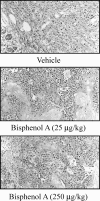
Bisphenol A (BPA) is an industrial plasticizer that leaches from food containers during normal usage, leading to human exposure. Early and chronic exposure to endocrine-disrupting environmental contaminants such as BPA elevates the potential for long-term health consequences. We examined the impact of BPA exposure on fetal programming of mammary tumor susceptibility as well as its growth promoting effects on transformed breast cancer cells in vivo. Fetal mice were exposed to 0, 25, or 250 μg/kg BPA by oral gavage of pregnant dams. Offspring were subsequently treated with the known mammary carcinogen, 7,12-dimethylbenz[a]anthracene (DMBA). While no significant differences in postnatal mammary development were observed, both low- and high-dose BPA cohorts had a statistically significant increase in susceptibility to DMBA-induced tumors compared to vehicle-treated controls. To determine if BPA also promotes established tumor growth, MCF-7 human breast cancer cells were subcutaneously injected into flanks of ovariectomized NCR nu/nu female mice treated with BPA, 17beta-estradiol, or placebo alone or combined with tamoxifen. Both estradiol- and BPA-treated cohorts formed tumors by 7 wk post-transplantation, while no tumors were detected in the placebo cohort. Tamoxifen reversed the effects of estradiol and BPA. We conclude that BPA may increase mammary tumorigenesis through at least two mechanisms: molecular alteration of fetal glands without associated morphological changes and direct promotion of estrogen-dependent tumor cell growth. Both results indicate that exposure to BPA during various biological states increases the risk of developing mammary cancer in mice.
Figures


Similar articles
-
In utero exposure to bisphenol A shifts the window of susceptibility for mammary carcinogenesis in the rat.Environ Health Perspect. 2010 Nov;118(11):1614-9. doi: 10.1289/ehp.1002148. Environ Health Perspect. 2010. PMID: 20675265 Free PMC article.
-
In utero exposure to diethylstilbestrol (DES) or bisphenol-A (BPA) increases EZH2 expression in the mammary gland: an epigenetic mechanism linking endocrine disruptors to breast cancer.Horm Cancer. 2010 Jun;1(3):146-55. doi: 10.1007/s12672-010-0015-9. Horm Cancer. 2010. PMID: 21761357 Free PMC article.
-
Prenatal bisphenol A exposure induces preneoplastic lesions in the mammary gland in Wistar rats.Environ Health Perspect. 2007 Jan;115(1):80-6. doi: 10.1289/ehp.9282. Environ Health Perspect. 2007. PMID: 17366824 Free PMC article.
-
Estrogens in the wrong place at the wrong time: Fetal BPA exposure and mammary cancer.Reprod Toxicol. 2015 Jul;54:58-65. doi: 10.1016/j.reprotox.2014.09.012. Epub 2014 Sep 30. Reprod Toxicol. 2015. PMID: 25277313 Free PMC article. Review.
-
Large effects from small exposures. III. Endocrine mechanisms mediating effects of bisphenol A at levels of human exposure.Endocrinology. 2006 Jun;147(6 Suppl):S56-69. doi: 10.1210/en.2005-1159. Epub 2006 May 11. Endocrinology. 2006. PMID: 16690810 Review.
Cited by
-
High levels of dietary soy decrease mammary tumor latency and increase incidence in MTB-IGFIR transgenic mice.BMC Cancer. 2015 Feb 6;15:37. doi: 10.1186/s12885-015-1037-z. BMC Cancer. 2015. PMID: 25655427 Free PMC article.
-
Molecular mechanism(s) of endocrine-disrupting chemicals and their potent oestrogenicity in diverse cells and tissues that express oestrogen receptors.J Cell Mol Med. 2013 Jan;17(1):1-11. doi: 10.1111/j.1582-4934.2012.01649.x. Epub 2012 Dec 20. J Cell Mol Med. 2013. PMID: 23279634 Free PMC article. Review.
-
Varying Susceptibility of the Female Mammary Gland to In Utero Windows of BPA Exposure.Endocrinology. 2017 Oct 1;158(10):3435-3447. doi: 10.1210/en.2017-00116. Endocrinology. 2017. PMID: 28938483 Free PMC article.
-
Hormones and endocrine-disrupting chemicals: low-dose effects and nonmonotonic dose responses.Endocr Rev. 2012 Jun;33(3):378-455. doi: 10.1210/er.2011-1050. Epub 2012 Mar 14. Endocr Rev. 2012. PMID: 22419778 Free PMC article. Review.
-
Bioremediation of phenolic pollutant bisphenol A using optimized reverse micelles system of Trametes versicolor laccase in non-aqueous environment.3 Biotech. 2021 Jun;11(6):297. doi: 10.1007/s13205-021-02842-4. Epub 2021 May 25. 3 Biotech. 2021. PMID: 34136334 Free PMC article.
References
-
- Maffini MV, Rubin BS, Sonnenschein C, Soto AM. Endocrine disruptors and reproductive health: the case of bisphenol-A. Mol Cell Endocrinol 2006; 254–255: 179 186 - PubMed
-
- Vandenberg LN, Hauser R, Marcus M, Olea N, Welshons WV. Human exposure to bisphenol A (BPA). Reprod Toxicol 2007; 24: 139 177 - PubMed
-
- Welshons WV, Nagel SC, vom Saal FS. Large effects from small exposures. III. Endocrine mechanisms mediating effects of bisphenol A at levels of human exposure. Endocrinology 2006; 147: S56 S69 - PubMed
-
- Goodson A, Robin H, Summerfield W, Cooper I. Migration of bisphenol A from can coatings—effects of damage, storage conditions and heating. Food Addit Contam 2004; 21: 1015 1026 - PubMed
Publication types
MeSH terms
Substances
Grants and funding
LinkOut - more resources
Full Text Sources
Other Literature Sources
Research Materials

