. 2011 Jul 22;109(3):272-80.
doi: 10.1161/CIRCRESAHA.111.240820. Epub 2011 Jun 2.

Placental growth factor regulates cardiac adaptation and hypertrophy through a paracrine mechanism

Affiliations

Placental growth factor regulates cardiac adaptation and hypertrophy through a paracrine mechanism

Federica Accornero et al. Circ Res. .

Abstract

Rationale: Paracrine growth factor-mediated crosstalk between cardiac myocytes and nonmyocytes in the heart is critical for programming adaptive cardiac hypertrophy in which myocyte size, capillary density, and the extracellular matrix function coordinately.

Objective: To examine the role that placental growth factor (PGF) plays in the heart as a paracrine regulator of cardiac adaptation to stress stimulation.

Methods and results: PGF is induced in the heart after pressure-overload stimulation, where it is expressed in both myocytes and nonmyocytes. We generated cardiac-specific and adult inducible PGF-overexpressing transgenic mice and analyzed Pgf(-/-) mice to examine the role that this factor plays in cardiac disease and paracrine signaling. Although PGF transgenic mice did not have a baseline phenotype or a change in capillary density, they did exhibit a greater cardiac hypertrophic response, a greater increase in capillary density, and increased fibroblast content in the heart in response to pressure-overload stimulation. PGF transgenic mice showed a more adaptive type of cardiac growth that was protective against signs of failure with pressure overload and neuroendocrine stimulation. Antithetically, Pgf(-/-) mice rapidly died of heart failure within 1 week of pressure overload, they showed an inability to upregulate angiogenesis, and they showed significantly less fibroblast activity in the heart. Mechanistically, we show that PGF does not have a direct effect on cardiomyocytes but works through endothelial cells and fibroblasts by inducing capillary growth and fibroblast proliferation, which secondarily support greater cardiac hypertrophy through intermediate paracrine growth factors such as interleukin-6.

Conclusions: PGF is a secreted factor that supports hypertrophy and cardiac function during pressure overload by affecting endothelial cells and fibroblasts that in turn stimulate and support the myocytes through additional paracrine factors.


Figures

Figure 1
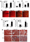
Figure 1. Cardiac-specific inducible PGF transgenic mice
A, mRNA levels of PGF from adult myocytes isolated from heart of mice subjected to TAC or sham procedure, compared with total non-myocytes from the heart. *P< 0.05 vs Sham (N=3 experiments). B, ELISA assay of PGF expression in hearts from sham and TAC operated wildtype mice. Pgf−/− mouse hearts were a negative control. *P< 0.05 vs Sham (N=3 experiments). C, Schematic representation of the binary transgenic mouse system. DTG refers to double transgenic. D, Western blot analysis of tTA control and PGF transgene expression in hearts from DTG mice at 0, 2, 4 and 8 weeks after the removal of Dox. E, ELISA quantification of PGF overexpression in hearts of tTA controls and PGF DTG mice at 4 and 8 weeks off Dox. *P< 0.05 vs tTA (N=3 experiments). F, ELISA quantification of PGF secretion in isolated myocytes and non-myocytes from hearts of mice subjected to TAC or sham procedures to compare against myocytes from DTG hearts. *P< 0.05 vs Sham; #P<0.05 vs TAC (N=3 experiments) G, Immunofluorescence staining in cardiac histological sections for PGF (green) counterstained with wheat germ agglutinin-TRITC (red) to outline cardiomyocytes in the indicated genotypes. Magnification is 200X
Figure 2
Figure 2. PGF DTG mice develop greater cardiac hypertrophy after TAC
A, Ventricular weight to body weight ratio (VW/BW) from tTA controls and PGF DTG mice subjected to 2 weeks of TAC or sham surgery. *P< 0.05 vs. Sham; # P< 0.05 vs. tTA TAC. B, Echocardiographic analysis of left ventricle posterior wall (LVPW) thickness in tTA controls and PGF DTG mice after 2 weeks of TAC or a sham surgery. *P< 0.05 vs Sham; #P< 0.05 vs tTA TAC. C, Length measurements D, width measurements E, total area measurements and F, length/width ratios from isolated adult cardiomyocytes from tTA control and PGF DTG hearts in mice subjected to 2 weeks of TAC. *P< 0.05 vs tTA. Number of cells analyzed is shown, summated from 3 mouse hearts in each group. G, Fractional shortening (FS%) measured by echocardiography 2 weeks after TAC stimulation or sham surgery in the indicated groups of mice. H, FS% measured by echocardiography 12 weeks after TAC stimulation or sham surgery in tTA controls and PGF DTG mice. I, FS% measured by echocardiography 5 weeks after TAC stimulation with one week of Angiotensin II/phenylephrine infusion in tTA and PGF DTG mice. *P< 0.05 vs. Sham; # P< 0.05 vs. tTA TAC.
Figure 3
Figure 3. PGF DTG mice show mild increases in angiogenesis and fibroblasts
A, Quantification of number of capillaries per cardiomyocyte (vessels/myocyte) in cardiac histological sections from tTA control and PGF DTG mice after 2 weeks of TAC or a sham surgery. *P< 0.05 vs Sham; #P< 0.05 vs tTA TAC. Three fields were quantified per mouse heart. Number of mouse hearts used is shown in the figure. B and C, quantitation of mRNA levels for CD34 and CD117 in tTA and PGF DTG hearts after 3 days of TAC. *P< 0.05 vs tTA. D, Representative staining of histological sections from tTA and DTG mice after 3 days of TAC or sham surgery for vimentin (green) and α-actinin (red). Magnification is 200X. E, Quantification of the percentage of vimentin positive cells per field in tTA and DTG mouse hearts after 3 days of TAC or sham surgery. *P< 0.05 vs Sham; #P< 0.05 vs tTA TAC. Number of mouse hearts analyzed is shown, from which 3 fields were quantified each. F, Hydroxyproline (OH-proline) content in hearts from sham and TAC operated mice of the indicated genotypes or after 5 months of aging. *P< 0.05 vs Sham; #P< 0.05 vs tTA. Number of mouse hearts analyzed is shown. G, Masson’s trichrome histological sections for fibrosis (blue) in tTA control and PGF DTG mouse hearts after 2 weeks of TAC or sham surgery, and at 5 months of age. Magnification is 200X.
Figure 4
Figure 4. PGF is required for the compensatory response to pressure overload
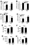
A, Ventricular weight to body weight ratio (VW/BW) in Wt controls and Pgf−/− mice subjected to 1 week of TAC or sham surgery. *P< 0.05 vs Sham; #P< 0.05 vs Wt TAC. Number of mice used is shown. B, Echocardiographic analysis of left ventricle posterior wall (LVPW) thickness after 1 week of TAC or sham surgery. *P< 0.05 vs Sham. C, Fractional shortening (FS%) measured by echocardiography 1 week after TAC or a sham surgery in Wt controls and Pgf−/− mice. *P< 0.05 vs Sham; #P< 0.05 vs Wt TAC. D, Echocardiographic analysis of left ventricular end diastolic dimension (LVED) in Wt controls and Pgf−/− mice after 1 week of TAC or a sham surgery. *P< 0.05 vs Sham; #P< 0.05 vs Wt TAC. E, Length measurements F, width measurements G, total area measurement and H, length/width ratio from adult cardiomyocytes isolated from hearts of Wt controls and Pgf−/− mice after 1 week of TAC. *P< 0.05 vs Wt. Number of cells analyzed is shown, summated from 3 mouse hearts in each group.
Figure 5
Figure 5. PGF is required for endothelial cells and fibroblasts response to pressure overload
A, Quantification of number of capillaries per cardiomyocyte (vessels/myocyte) in histological heart sections from Wt control and Pgf−/− mice after 1 week of TAC or a sham surgery. *P< 0.05 vs Sham; #P< 0.05 vs Wt TAC. Three fields were quantified per mouse heart. Number of mouse hearts used is shown in the figure. B and C, quantitation of mRNA levels for CD34 and CD117 in Wt and Pgf−/− hearts after 3 days of TAC. *P< 0.05 vs Wt. D, Representative staining of histological heart sections from Wt and Pgf−/− mice after 3 days of TAC or sham surgery by vimentin (green) and α-actinin (red). Magnification is 200X. E, Quantification of the percentage of vimentin positive fibroblasts per histological field in Wt and Pgf−/− hearts after the mice were subjected to 3 days of TAC or a sham surgery. *P< 0.05 vs Sham; #P< 0.05 vs Wt TAC. Number of mouse hearts analyzed is shown, from which 3 fields were quantified each.
Figure 6
Figure 6. PGF directly affects endothelial cells and cardiac fibroblasts
A, Representative pictures of HUVEC tube formation in Matrigel when co-cultured with AdβGal or AdPGF infected primary cardiomyocytes. B, Quantification of relative tube formation in HUVECs co-cultured with cardiomyocytes as described in A. *P< 0.05 vs AdβGal (N=3 experiments). C, Quantification of the proliferation rate of neonatal rat fibroblasts infected with AdβGal or AdPGF. *P< 0.05 vs AdβGal. (N=2 experiments, 8 wells each). D, Western blot for ERK1/2 phosphorylation in non-myocytes from the heart and isolated cardiomyocytes infected with AdbGal or AdPGF overnight. E, Quantification of surface area in neonatal rat cardiomyocytes infected with AdβGal or AdPGF (white bars) or treated for 24 hrs with conditioned media from fibroblasts infected with either adenovirus (black bars). *P< 0.05 vs no conditioned media; #P<0.05 vs AdβGal conditioned media. At least 50 cells were measured separately in 2 independent experiments. F, qRT-PCR results for the indicated growth factors from cardiac fibroblasts infected with AdPGF or AdβGal. *P<0.05 vs AdβGal. G,H, and I show qRT-PCR for the indicated mRNAs from hearts of the indicated mice after sham of 3 days of TAC stimulation. At least 3 mice were analyzed for each condition. *P<0.05 versus sham; #P<0.05 vs tTA or Wt TAC.

References

    1. Hunter JJ, Chien KR. Signaling pathways for cardiac hypertrophy and failure. N Engl J Med. 1999;341:1276–1283. - PubMed
    1. Lorell BH, Carabello BA. Left ventricular hypertrophy: Pathogenesis, detection, and prognosis. Circulation. 2000;102:470–479. - PubMed
    1. Frey N, Olson EN. Cardiac hypertrophy: The good, the bad, and the ugly. Annu Rev Physiol. 2003;65:45–79. - PubMed
    1. Haq S, Choukroun G, Lim H, Tymitz KM, del Monte F, Gwathmey J, Grazette L, Michael A, Hajjar R, Force T, Molkentin JD. Differential activation of signal transduction pathways in human hearts with hypertrophy versus advanced heart failure. Circulation. 2001;103:670–677. - PubMed
    1. Dorn GW, 2nd, Force T. Protein kinase cascades in the regulation of cardiac hypertrophy. J Clin Invest. 2005;115:527–537. - PMC - PubMed

Publication types

MeSH terms