. 2011 Dec;18(12):1904-13.
doi: 10.1038/cdd.2011.71. Epub 2011 Jun 3.

SAHA shows preferential cytotoxicity in mutant p53 cancer cells by destabilizing mutant p53 through inhibition of the HDAC6-Hsp90 chaperone axis

Affiliations

SAHA shows preferential cytotoxicity in mutant p53 cancer cells by destabilizing mutant p53 through inhibition of the HDAC6-Hsp90 chaperone axis

D Li et al. Cell Death Differ. 2011 Dec.

Abstract

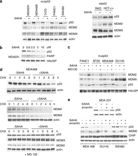
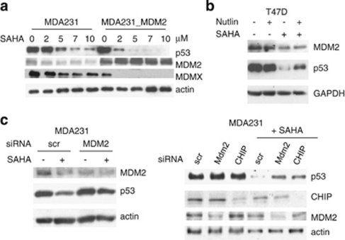
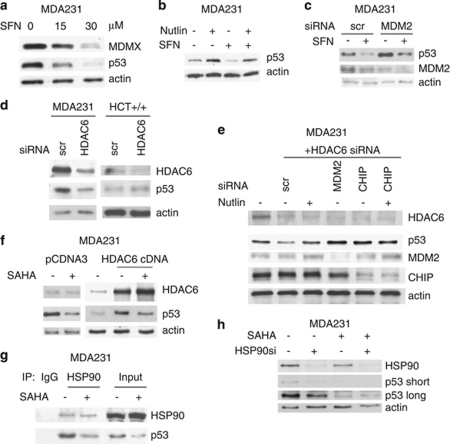
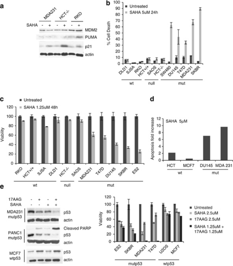
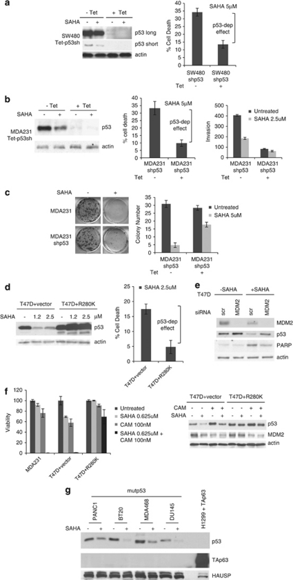
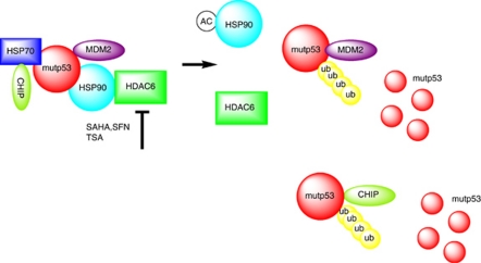
Mutant p53 (mutp53) cancers are surprisingly dependent on their hyperstable mutp53 protein for survival, identifying mutp53 as a potentially significant clinical target. However, exploration of effective small molecule therapies targeting mutp53 has barely begun. Mutp53 hyperstabilization, a hallmark of p53 mutation, is cancer cell-specific and due to massive upregulation of the HSP90 chaperone machinery during malignant transformation. We recently showed that stable complex formation between HSP90 and its mutp53 client inhibits E3 ligases MDM2 and CHIP, causing mutp53 stabilization. Histone deacetylase (HDAC) inhibitors (HDACi) are a new class of promising anti-cancer drugs, hyperacetylating histone and non-histone targets. Currently, suberoylanilide hydroxamic acid (SAHA) is the only FDA-approved HDACi. We show that SAHA exhibits preferential cytotoxicity for mutant, rather than wild-type and null p53 human cancer cells. Loss/gain-of-function experiments revealed that although able to exert multiple cellular effects, SAHA's cytotoxicity is caused to a significant degree by its ability to strongly destabilize mutp53 at the level of protein degradation. The underlying mechanism is SAHA's inhibition of HDAC6, an essential positive regulator of HSP90. This releases mutp53 and enables its MDM2- and CHIP-mediated degradation. SAHA also strongly chemosensitizes mutp53 cancer cells for chemotherapy due to its ability to degrade mutp53. This identifies a novel action of SAHA with the prospect of SAHA becoming a centerpiece in mutp53-specific anticancer strategies.


Figures

Figure 1
Figure 1
SAHA downregulates mutp53 but not wtp53. This effect occurs at the level of protein degradation but not at the level of transcription. (a) SAHA (5 μM) strongly downregulates mutant p53, MDM2 and MDMX protein levels. In contrast, SAHA does not alter levels of wild-type p53 protein. A panel of human tumor cell lines harboring mutant or wild-type p53 were analyzed by immunoblot as indicated. Actin, loading control. (b) SAHA-mediated downregulation of mutant p53 is dose-dependent and correlates with induction of PARP cleavage. (c) Proteosome inhibitor MG132 (5 μM) rescues SAHA-mediated downregulation of mutp53, MDM2 and MDMX. Cells were simultaneously treated with SAHA (5 μM) and MG132 (5 μM) for 16 h. HAUSP was used as a loading control. (d) SAHA (5 μM) dramatically decreases the half-life of mutant p53, MDM2 and MDMX, all bona fide physiologic substrates of MDM2. CHX chase for the indicated times. Actin, loading control. Bottom, proteasome inhibitor MG132 (5 μM) rescues SAHA-induced destabilization of mutp53, MDM2 and MDMX after CHX treatment. (e) The SAHA-mediated downregulation of mutant p53 does not occur at the level of transcription. Mutp53 cells were treated with SAHA (5 μM) and/or α-amanitin (10 μg/ml) for 16 h, a potent and specific transcriptional inhibitor of RNA polymerase II
Figure 2
Figure 2
SAHA-induced degradation of mutp53 is mediated by reactivation of MDM2 and CHIP E3 ligases. (a) Overexpression of MDM2 enhances SAHA-induced degradation of mutp53 and MDMX. The cell system used here is described by Li et al. wherein we show that in human cancer cells endogenous mutant p53 – despite its ability to interact with MDM2 – suffers from a profound lack of ubiquitination as the root cause of its degradation defect. Multiple lines of evidence indicate the functional impairment of MDM2 in mutp53 cancer cells by the HSP90 chaperone. We found that in contrast to transiently overexpressed MDM2, physiologically tolerated, stably overexpressed MDM2 is silent and fails to affect ubiquitination and mutp53 levels, indicating the presence of selective pressure against active MDM2 in mutp53 cancer cells. Immunoblot of parental MDA231 and cells stably overexpressing MDM2. Actin as loading control. (b) Nutlin partially prevents SAHA-induced destabilization of mutp53, indicating MDM2 reactivation upon SAHA (5 μM). GAPDH, loading control. (c) siRNA-mediated knockdown of MDM2 and CHIP partially rescues SAHA-induced (5 μM) destabilization of mutp53
Figure 3
Figure 3
Inhibition of HDAC6 inhibits HSP90 and destabilizes mutp53 by reactivating MDM2 and CHIP. (a) HDAC6 inhibitor SFN destabilizes mutp53 and MDMX. Nutlin (b) and MDM2 siRNA (c) partially prevent SFN-induced degradation of mutp53. (d) Likewise, HDAC6 inhibition by siRNA also destabilizes mutp53 (in MDA231) but does not destabilize wtp53 (in HCT116). (e) siRNA-mediated knockdown of HDAC6 results in degradation of mutp53 that is rescued by Nutlin (lane 3) or by siRNA against MDM2 and CHIP. Scr, scrambled siRNA. Immunoblot. (f) Conversely, HDAC6 overexpression stabilizes mutp53, which is significantly rescued by concomitant SAHA. Immunoblot. (g) SAHA (5 μM) inhibits the complex between Hsp90 and mutp53. Lysates from MDA231 cells left untreated or treated with SAHA were immunoprecipitated with IgG or anti-Hsp90. (h) In HSP90-ablated cells, SAHA does not induce further destabilization of mutp53. MDA231 cells were transfected with siHSP90 or scrambled siRNA control. After 24 h, cells were treated with 2.5 μM SAHA for an additional 24 h followed by immunoblot
Figure 4
Figure 4
SAHA shows preferential cytotoxicity for mutp53 tumor cells. (a) SAHA does not pharmacologically rescue mutant p53 to assume wild type function. MDM2 is not induced. Although p21 is slightly induced, it appears to be in a p53-independent manner, based on p53-deficient HCT116 cells (HCT−/−). SAHA was used at 5 μM. (bd) SAHA shows strong cytotoxic cell killing towards mutp53 tumor cells, as measured by trypan blue exclusion assay (b), cell viability CTB assay (c) and subG1 FACS (d). In contrast, wtp53 and p53 null tumor cells show only a minimal cytotoxic response to SAHA. Random panel of human tumor cells. (e) SAHA and 17AAG can synergize to induce preferential apoptosis of mutp53 cancer cells. Co-treatment of SAHA and 17AAG causes a synergistic loss of cell viability specifically in MDA231 and T47D. Combined efficacy correlates with the degree of mutp53 destabilization and PARP cleavage (right immunoblots). This synergism is due to complementary drug targets within the HSP90 chaperone machinery
Figure 5
Figure 5
Causality – SAHA's preferential cytotoxic effect on mutp53-harboring cancer cells is to a significant degree due to its ability to degrade mutp53. (ac) Pseudo-null lines were generated to eliminate mutp53 as a target of SAHA. Cells were pretreated with tetracycline to achieve ‘isogenicity'. Cells with high (control) and very low levels of mutp53 were then treated with SAHA. In both MDA231 and SW480, SAHA partially loses cytotoxicity when mutp53 is very low. (a and b) In mutp53-harboring cancer cells SAHA loses over 50% killing efficacy when mutp53 is knocked down. Left, immunoblots of (a) SW480 and (b) MDA231 cells stably harboring Tet-inducible shp53. Middle, trypan blue exclusion assays. Percent increase in cell death of SAHA-treated cells relative to their respective untreated controls without SAHA. (b, right) Knockdown of mutp53 by SAHA or/and Tet- inducible shRNA inhibits invasion. (b, left) Inhibition of invasion directly correlates with the extent of mutp53 destabilization. Aliquots were used for the Matrigel Boyden chambers and corresponding immunoblot. (c) Clonogenicity assay of MDA231 cells harboring Tet-inducible shp53 in response to SAHA. Right, quantitation. For experiments in a–c, cells were pretreated with tetracycline for 48 h and then seeded for subsequent SAHA treatment (5 μM) for an additional 24 h. (d) Likewise, SAHA loses killing efficacy when its ability to degrade mutp53 is overwhelmed by excess ectopic mutant p53. Left, immunoblots of empty vector or mutp53 R280K overexpressing T47D. Right, increase in cell death of SAHA-treated cells relative to their respective untreated controls without SAHA. Trypan blue exclusion assay. (e) siRNA-mediated knockdown of MDM2 partially rescues SAHA-induced destabilization of mutp53 and inhibits the cytotoxic effect of SAHA, indicated by reduced PARP cleavage. Cells were transfected with siMDM2 or scrambled siRNA, followed by SAHA treatment (5 μM) for 16 h. Immunoblot, actin as loading control. (f) SAHA strongly chemosensitizes mutp53 cancer cells and this is due to its ability to degrade mutp53. Left, viability of MDA231, T47D vector and T47D cells overexpressing mutp53 R280K (see Figure 5d) after low-dose camptothecin (100 nM) and SAHA (625 nM) treatment alone and in combination. CTB assays. Right, corresponding immunoblots of the T47D set. (g) SAHA fails to induce TAp63 protein in mutant p53 cancer cells. Despite multiple forced attempts (overloading, overexposure), we were unable to detect SAHA-induced TAp63 protein levels in any of the cell lines. Immunoblot with a pan-p63 antibody (H137) of total cell lysates (20 μg per lane) from mutp53 cancer cells grown in the absence or presence of SAHA (5 μM) for 24 h. As control, 2 μg of total cell lysate from H1299 cells transfected with a TAp63α plasmid was loaded
Figure 6
Figure 6
Proposed model of SAHA-mediated destabilization of mutant p53 by inhibiting the HDAC6-Hsp90 chaperone axis. Based on results from us and others, we propose the following model. Normal tissues that harbor missense mutp53 are able to efficiently control their mutp53 levels despite the fact that their MDM2 levels are only supported by constitutive P1 promoter-derived transcription. In contrast, tumor-specific stabilization of mutp53 proteins, which contributes to driving the tumor phenotype, depends on a second alteration these cells undergo upon malignant transformation. This is their addiction to support from the activated heat shock machinery for survival. In contrast to wtp53, the aberrant conformation of mutp53 proteins requires permanent heat shock support, thus mutp53 is stably engaged in complexes with the highly activated HSP90 chaperone to prevent aggregation. Intimately linked to this conformational stabilization, however, is the fact that this interaction also acts as a large protective ‘cage' against degradation, thereby enabling mutp53's GOF. The E3 ligases MDM2 and CHIP, which in principle are capable of degrading mutp53, might also be trapped in this complex in an inactive state. As mutp53 is fully competent to bind to MDM2, HSP90 likely binds to pre-existing mutp53-MDM2 complexes. Alternatively, chaperone-bound mutp53 could recruit MDM2. HDAC6 is a cytoplasmic non-histone HDAC that deacetylates Hsp90 and an obligate positive regulator of the HSP90 chaperone activity., Inhibiting HDAC6 by SAHA and related drugs leads to hyperacetylation and inhibition of Hsp90. This destroys the complex, releases mutp53 and enables MDM2/CHIP-mediated degradation

References

    1. Lang GA, Iwakuma T, Suh YA, Liu G, Rao VA, Parant JM, et al. Gain of function of a p53 hot spot mutation in a mouse model of Li-Fraumeni syndrome. Cell. 2004;119:861–872. - PubMed
    1. Olive KP, Tuveson DA, Ruhe ZC, Yin B, Willis NA, Bronson RT, et al. Mutant p53 gain of function in two mouse models of Li-Fraumeni syndrome. Cell. 2004;119:847–860. - PubMed
    1. Blandino G, Levine AJ, Oren M. Mutant p53 gain of function: differential effects of different p53 mutants on resistance of cultured cells to chemotherapy. Oncogene. 1999;18:477–485. - PubMed
    1. Li R, Sutphin PD, Schwartz D, Matas D, Almog N, Wolkowicz R, et al. Mutant p53 protein expression interferes with p53-independent apoptotic pathways. Oncogene. 1998;16:3269–3277. - PubMed
    1. Bossi G, Lapi E, Strano S, Rinaldo C, Blandino G, Sacchi A. Mutant p53 gain of function: reduction of tumor malignancy of human cancer cell lines through abrogation of mutant p53 expression. Oncogene. 2006;25:304–309. - PubMed

Publication types

MeSH terms