. 2011;6(5):e20540.
doi: 10.1371/journal.pone.0020540. Epub 2011 May 26.

Vascular wall-resident CD44+ multipotent stem cells give rise to pericytes and smooth muscle cells and contribute to new vessel maturation

Affiliations

Vascular wall-resident CD44+ multipotent stem cells give rise to pericytes and smooth muscle cells and contribute to new vessel maturation

Diana Klein et al. PLoS One. 2011.

Abstract

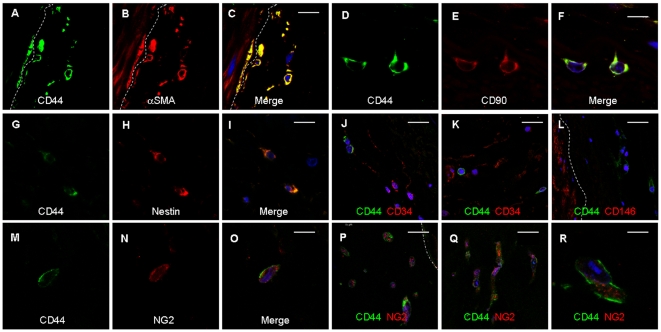
Here, we identify CD44(+)CD90(+)CD73(+)CD34(-)CD45(-) cells within the adult human arterial adventitia with properties of multipotency which were named vascular wall-resident multipotent stem cells (VW-MPSCs). VW-MPSCs exhibit typical mesenchymal stem cell characteristics including cell surface markers in immunostaining and flow cytometric analyses, and differentiation into adipocytes, chondrocytes and osteocytes under culture conditions. Particularly, TGFß1 stimulation up-regulates smooth muscle cell markers in VW-MPSCs. Using fluorescent cell labelling and co-localisation studies we show that VW-MPSCs differentiate to pericytes/smooth muscle cells which cover the wall of newly formed endothelial capillary-like structures in vitro. Co-implantation of EGFP-labelled VW-MPSCs and human umbilical vein endothelial cells into SCID mice subcutaneously via Matrigel results in new vessels formation which were covered by pericyte- or smooth muscle-like cells generated from implanted VW-MPSCs. Our results suggest that VW-MPSCs are of relevance for vascular morphogenesis, repair and self-renewal of vascular wall cells and for local capacity of neovascularization in disease processes.


Conflict of interest statement

Competing Interests: The authors have declared that no competing interests exist.

Figures

Figure 1
Figure 1. Putative MSCs within the hITA wall.
(A) Immunostainings of hITA sections show that αSMA (alpha smooth muscle actin) is mainly detected in SMC, but also in single cells of vasculogenic zone (arrows). (B) CD146 staining is limited to the SMC layer. (C–F) CD73 and RGS5 (regulator of G-protein signaling 5) show the same staining patterns as αSMA. (G–J) CD90 and nestin positive cells are seen in the vasculogenic zone within the adventitia (arrows). (K) CD44 staining is only found in single cells within the adventitia, prominently near to the media (L, M) as visualized by higher magnification (arrows). (N) Sox2 staining is also found in single cells within the adventitia. (O, P) After performing ring assay, when small hITA sections were embedded in GFR-Matrigel and cultured for 2–7 days, after 2–3 days numerous cells positive for both CD44 and NG2 are detectable in Matrigel (arrows) indicating the mobilization and sprouting capacity of the CD44+ putative MSCs from the hITA wall. Lu lumen, TM tunica media, Ad adventitia, MG Matrigel. Dotted line marks the border between media and adventitia of the hITA wall. Bar A–C, E, G, I, K 50 µm; D, F, H, J, M, N 10 µm, L, P 25 µm.
Figure 2
Figure 2. Co-localisation of MSC marker proteins in CD44+ cells within the hITA wall.
Double-immunostainings of hITA sections using antibodies against typical MSC maker proteins and CD44 demonstrate that CD44+ cells within vasculogenic zone closed to tunica media (emphasised by dashed line) are also positive for αSMA (A–C), CD90 (D–F), and nestin (G–I), but they are negative for endothelial and hematopoietic progenitor cell marker CD34 (J–K), and lack also CD146 expression (L). Single CD44+ cells within that zone express NG2 (N–O) and these cells migrate into the outside of the vessel wall when performing ring assay studies (P–R; blue, TOTO®-3 iodide).
Figure 3
Figure 3. Improved purification and characterisation of vascular wall-derived MPSCs.
(A) FACS analysis on MPSCs isolated from hITA wall and selected by MACS using CD44 monoclonal antibody show that they are positive for CD90, CD73, CD105, CD44, Stro1 and CD29 but negative for lineage cell markers CD45, CD68, CD11b, CD19, SMC markers CD146 and PDGFRβ and endothelial cell markers CD34, KDR and CD31 indicating no considerable contamination by other types of progenitors. FACS data representative for at least 3 independent experiments with similar results are shown. (B) Cultivated vascular wall-derived CD44+ cells show flattened and fibroblast-like morphology and form clonally cell aggregates upon prolonged culturing (CFU, colony forming units). Bar 50 µm. Immunofluorescent analysis on cultivated CD44+ MPSCs shows that they are also positive for RGS5 (green) and express stem cell marker Sox2 (green) and Oct4 (red) using confocal microscopy (blue, TOTO®-3 iodide). Bar, 15 µm.
Figure 4
Figure 4. Exogenous TGFβ1 reduces in-gel sprouting of cultured vascular wall-derived MPSCs.
Vascular wall-derived MPSCs were embedded in GFR-Matrigel as 3D-spheroids and exposed to normal growth media (NGM), supernatant of the tumor cell lines PC3 and A549 and indicated factors. (A) VW-MPSCs in-gel sprouting and Matrigel invasion was observed by phase contrast microscopy, and (B) quantified after 48 hours of stimulation as shown in the diagram. Application of PC3 and A549 supernatant and FGF2 increases the capacity of cell invasion in the Matrigel while the presence of TGFß1 alone or in combination with indicated factors suppresses it. The data represent the mean cumulative length of all cord-like sprouts growing from 10 individual spheroids per experimental group. The figure shows the results from 1 of 4 independent experiments with similar results. *, p<0.05; **, p≤0.005.
Figure 5
Figure 5. Pericyte-like coverage of endothelial tube-like structures by vascular wall-derived MPSCs in vitro.
Isolated vascular wall-derived MPSCs (labelled red) are cultured alone or together with HUVEC (human umbilical cord endothelial cells; labelled green) on Matrigel. VW-MPSCs alone form cord-like structures (A) but VW-MPSCs together with HUVEC (B–I) form more prominent network suggesting capillary-like structures in phase contrast microscopy (B). Confocal microscopy analysis after 5–7 hours (C) and after 5 days (D–I) shows a tight association of VW-MPSCs to the capillary-like EC (D). Under additional application of VEGF-A (10 ng/ml) HUVEC form prominent capillary-like structures (green) which are covered by VW-MPSCs (red) (E–I). Higher magnification of these structures reveals a pericyte-like assembly (arrows) of VW-MPSCs to the tube like structures (F–I). Tight association of VW-MPSCs to the capillary-like EC can nicely be seen by combining fluorescence and phase contrast microscopy (H, I). Photographs representative for at least 3 independent experiments with similar results are shown. Bar A–C 50 µm; D,E, 100 µm, F–H15 µm, I 5 µm.
Figure 6
Figure 6. Differential expressions of marker genes in vascular wall-derived MPSCs versus SMC.
(A) QRT-PCR analyses show that genes specific for SMC such as alpha smooth muscle actin (αSMA, ACTA2), TAGLN1 (transgelin), THSP1 (Thrombospondin 1), MYOC (myocardin) and HPLN1 hyaluronan and proteoglycan link protein 1 are expressed significantly higher in hAoSMC (human aortic smooth muscle cells) in comparison to MSCs while PDGFRα (platelet-derived growth factor α) and NG2 are expressed stronger in vascular wall-derived MPSCs (A–B). Stimulation of VW-MPSCs with VEGF165, PDGF-BB, FGF2 (10 ng/ml), TGFβ1 (5 ng/ml) alone or in indicated combinations for 14 days shows an up-regulation of SMC markers TAGLN and THSP1 as compared to VW-MPSCs cultured in normal growth media (NGM) (C). Resulting expression levels were normalized by division through the mean expression value of the reference gene (β-actin). Data are presented as mean ± SD from four independent experiments measured at least two times each. *, p<0.05; **, p≤0.005. The stimulation of VW-MPSCs by TGFß1 alone or in combination VEGF and PDGF also increases the protein level of SMC markers αSMA and TAGLN as shown by immunoblotting (D). Total cell lysates were generated by scraping cells in ice-cold RIPA buffer. Equal protein amounts were subjected for SDS-PAGE. TAGLN was detected by Western blot using chemiluminescence. β-actin was included as a loading control. Data representative for at least 3 independent experiments with similar results are shown.
Figure 7
Figure 7. Contribution of vascular wall-derived MPSCs to new vessel formation in vivo.
VW-MPSCs and HUVEC were grafted into Scid mice subcutaneously for 14 days as spheroids in Matrigel supplemented with growth factors. Immunofluorescent analysis of isolated plug tissues was performed. Double-staining for hCD34 (green) and αSMA (red) shows a close assembly of αSMA+ cells to the vessel wall formed by HUVEC (A, C).Functionally perfused blood vessels within the plugs are identified by presence of erythrocytes within the vessel lumen as detected by phase contrast microscopy (B, arrow). The specificity of hCD34 was demonstrated by its absence in blood vessels of normal mouse fatty tissue (D). Furthermore, double stainings for hCD34 (red) and TAGLN (green) (E–I) show flattened TAGLN+ positive cells in tight association to vessels formed by implanted HUVEC within the plugs (E–G). Intensive vascularisation of plugs is also seen when VW-MPSCs/HUVEC are grafted in Matrigel together with VEGF and FGF2 (A–F, I) as well as TGFβ1 alone (G, H). Cells strongly positive for TAGLN surround tightly the vessels formed by HUVEC (G, arrowheads) while also some single roundly shaped and TAGLN negative cells (arrow) are present (G, arrow). Few flattened and TAGLN+ cells (arrowhead) which are not assembled to new vessels are found in Matrigel indicating the presence of SMC, while some rounded cells (arrowheads) are only weak positive for TAGLN which probably represent still differentiating MPSCs (H). EGFP-labeling of VW-MPSCs shows co-localization of TAGLN (red) and EGFP fluorescence identifying the EGFP-labeled VW-MPSCs as the source of the pericytes and SMC-like cells surrounding the vessels (blue, TOTO®-3 iodide) (I). Bar A, C 20 µm; E, H, I 10 µm; B, F, G 5 µm. Electron microscopic analysis demonstrates a capillary with endothelial cells (EC) and regularly assembled pericytes (PC) covering endothelial cells. The presence of erythrocytes (Ery) within the capillary lumen indicates the connection of this capillary to the blood perfusion (J). In some areas immature vessels are seen as observed by EC morphology and absence of pericytes in the vessel wall (K). Also single cells or small cell clusters with contractile filaments in the cytoplasm are found indicating the presence of SMC, probably generated from the implanted SMCs as shown by higher magnification (L).

References

    1. Carmeliet P, Jain RK. Angiogenesis in cancer and other diseases. Nature. 2000;407:249–257. - PubMed
    1. Ferrara N, Kerbel RS. Angiogenesis as a therapeutic target. Nature. 2005;438:967–974. - PubMed
    1. Asahara T, Murohara T, Sullivan A, Silver M, van der Zee R, et al. Isolation of putative progenitor endothelial cells for angiogenesis. Science. 1997;275:964–967. - PubMed
    1. Prokopi M, Pula G, Mayr U, Devue C, Gallagher J, et al. Proteomic analysis reveals presence of platelet microparticles in endothelial progenitor cell cultures. Blood. 2009;114:723–32. - PubMed
    1. Aicher A, Zeiher AM, Dimmeler S. Mobilizing endothelial progenitor cells. Hypertension. 2005;45:321–325. - PubMed

Publication types

MeSH terms