. 2011;6(5):e20461.
doi: 10.1371/journal.pone.0020461. Epub 2011 May 26.

ECAT11/L1td1 is enriched in ESCs and rapidly activated during iPSC generation, but it is dispensable for the maintenance and induction of pluripotency

Affiliations

ECAT11/L1td1 is enriched in ESCs and rapidly activated during iPSC generation, but it is dispensable for the maintenance and induction of pluripotency

Kumiko A Iwabuchi et al. PLoS One. 2011.

Abstract

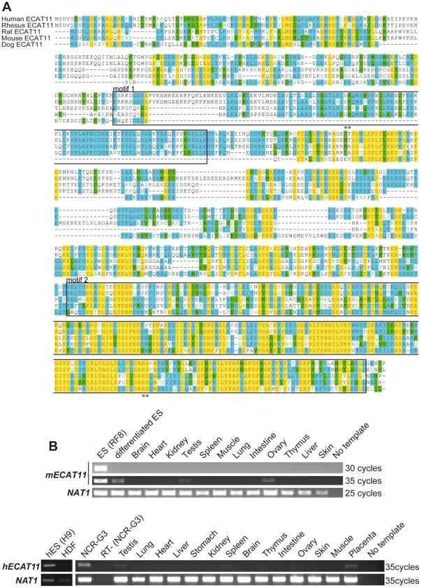
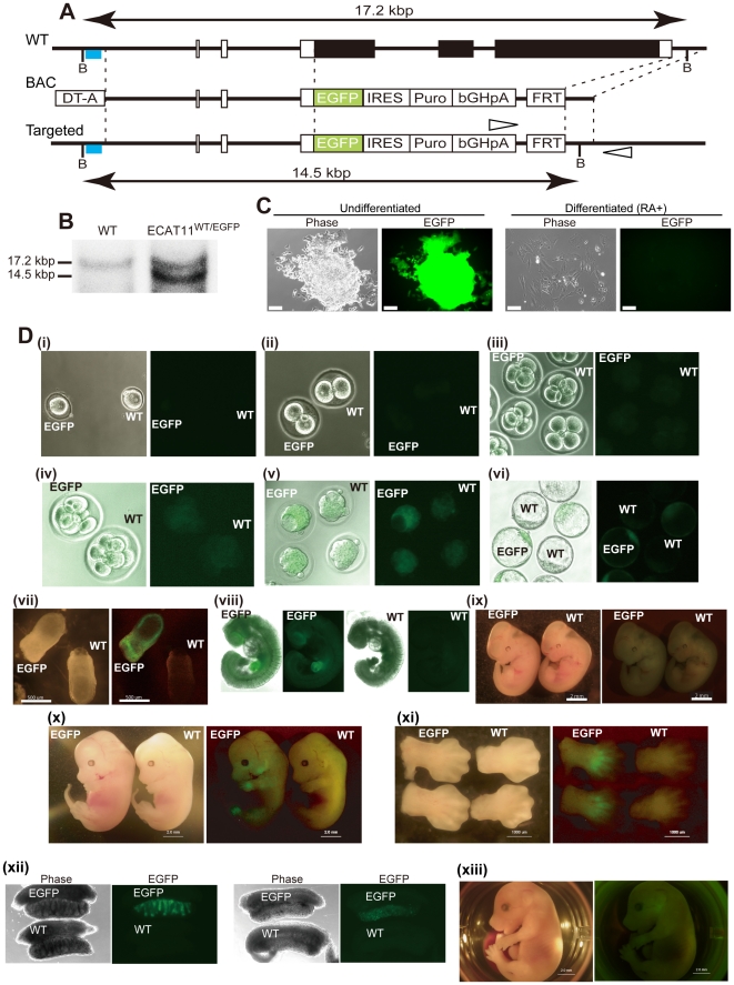
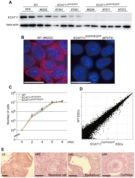
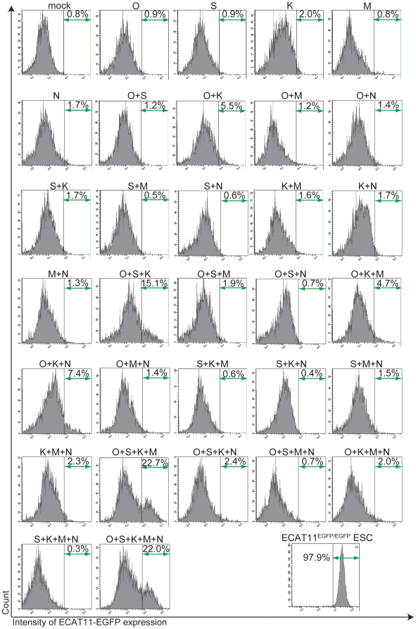
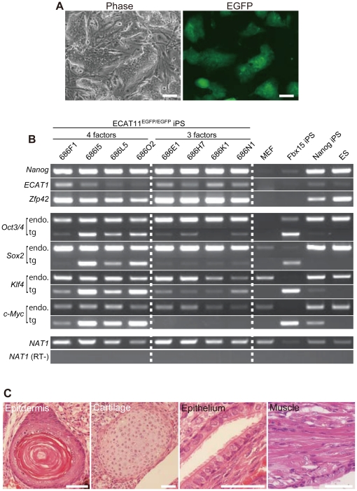
The principal factors that lead to proliferation and pluripotency in embryonic stem cells (ESCs) have been vigorously investigated. However, the global network of factors and their full signaling cascade is still unclear. In this study, we found that ECAT11 (L1td1) is one of the ESC-associated transcripts harboring a truncated fragment of ORF-1, a component of the L1 retrotransposable element. We generated an ECAT11 knock-in mouse by replacing its coding region with green fluorescent protein. In the early stage of development, the fluorescence was observed at the inner cell mass of blastocysts and epiblasts. Despite this specific expression, ECAT11-null mice grow normally and are fertile. In addition, ECAT11 was dispensable for both the proliferation and pluripotency of ESCs.We found rapid and robust activation of ECAT11 in fibroblasts after the forced expression of transcription factors that can give rise pluripotency in somatic cells. However, iPS cells could be established from ECAT11-null fibroblasts. Our data demonstrate the dispensability of ECAT11/L1td1 in pluripotency, despite its specific expression.


Conflict of interest statement

Competing Interests: The authors have declared that no competing interests exist.

Figures

Figure 1
Figure 1. Protein structure andexpression of ECAT11.
(A) Amino acid sequences of ECAT11 from various species. The boxes indicate the conserved Transposase_22 motif. Double asterisks indicate equivalent regions of arginin residues responsible for the RNA binding activity of L1ORF1. Black letters on a white background; non-similar residues, blue letters on a cyan background; consensus residues derived from a block of similar residues at a given position, black letters on a green background; consensus residues derived from the occurrence of >50% of a single residue at a given position, red letters on a yellow background; consensus residues derived from a completely conserved residue at a given position, green letters on a white background; residues weakly similar to the consensus residue at a given position. (B)The expression profiles of mouse (upper)and human (lower) ECAT11. RNA was isolated from ESCs, differentiated ESCs, human ESCs (H9), human dermal fibroblasts (HDF), an embryonic tumor cell line (NCR-G3)and various tissues from adult mice or humans, and were used for an RT-PCR analysis. The differentiation of mouse ESCs was achieved by retinoic acid (RA) treatment (300 nM) for 7 days. The amplification cycles are shown at right. NAT1 was used as a loading control.
Figure 2
Figure 2. Generation of the ECAT11-EGFP knock-in reporter and mice.
(A) Schematic representations of the structures of the mouse ECAT11 gene (WT), targeting BAC (BAC), and targeted loci (Targeted). The positions of the probes (blue lines), and recognition sites of BspHI (B) are shown. The black box indicates the ECAT11 coding region. Open arrowheads indicate the 3′ screening primer pair that amplified only the recombinant allele. DT-A: diphtheria toxin A, IRES: internal ribosomal entry site, Puro: puromycin resistance gene, bGHpA: bovine growth hormone poly A sequence, FRT: flippase recognition target. (B) The results of the Southern blot analysis of ECAT11WT/EGFP ESCs. (C)The morphology of the ECAT11EGFP/EGFP ESCs grown on gelatin-coated dishes. The phase and EGFP fluorescence images of undifferentiated (left panels) and differentiated (right panels) cells are shown. Cells were cultured with RA for eight days before taking the photographs. Bars; 100 µm. (D) ECAT11-EGFP expression in mouse embryos. Bright field and EGFP fluorescence images of fertilized eggs (i), 2 cells (ii), 4 cells (iii), 8 cells (iv), morulae (v), blastocysts (vi), E7.5 gastrulae (vii), E9.5 embryos (viii), E10.5 embryos (ix), E13.5 embryos (x), limbs of E13.5 embryos (xi), reproductive glands of E13.5 embryos; left, testis; right, ovary (xii), and the E15.5 embryo of an ECATEGFP/EGFP mouse (xiii) are shown. WT: wild type. EGFP: ECAT11EGFP/EGFP.
Figure 3
Figure 3. ECAT11 is dispensable for mouse ESCs.
(A)A Western blot analysis of the ECAT11 expression in ESC lines. The data are shown in duplicate. (B) Immunocytochemistry of ECAT11. Red: ECAT11, Blue: DAPI. Bars; 20 µm. (C)The proliferation of ECAT11 knock-in ESCs. Ten thousand cells were plated, and counted on days2, 4, 6 and 8. We used 2 WT, 2 ECAT11WT/EGFP, and 3 ECAT11EGFP/EGFP ESC lines for this analysis. The data are shown as the averagesand standard deviations. (D) Scatter plots showing a comparison of the global gene expression determined by the microarray analyses between the WT and ECAT11EGFP/EGFP ESCs. The data from 2 WT ESC lines and 3 ECAT11EGFP/EGFP ESC lines cultured on gelatin coated dishes were averaged and used for this analysis. (E)Hematoxylin and eosin staining of teratomas generated from ECAT11EGFP/EGFP ESCs. The gross image (upper left), neural tissue, gut-like epithelia, and cartilage are shown. The scale bars are 200 µm in the gross image and 100 µm in the other images.
Figure 4
Figure 4. Activation of the ECAT11 promoter by forced expression of Oct4, Sox2 and Klf4.
The expression of the ECAT11-EGFP fluorescent marker was induced by various combinations of transcription factors. EGFP fluorescence was analyzed by flowcytometry two days after retroviral transduction. O: Oct4, S: Sox2, K: Klf4, M: c-Myc, N: Nanog.
Figure 5
Figure 5. Generation of iPSCs from ECAT11EGFP/EGFP MEFs.
(A) Morphology of an ECAT11EGFP/EGFP iPSC colony, which was picked on day 23 after induction of four factors (Oct4, Sox2, Klf4 and c-Myc) and cultured on feeder cells for three passages. Scale bars: 100 µm. (B) The expression levels of three pluripotency markers (Nanog, ECAT1 and Zfp42), and four transcription factors (Oct4, Sox2, Klf4 and c-Myc). Total RNA was collected from four clones of ECAT11EGFP/EGFP iPSCs(686F1, 686I5, 686L5 and 686O2) established using four factors (Oct4, Sox2, Klf4 and c-Myc), and four clones (686E1, 686H7, 686K1 and 686N1)established usingthree factors (Oct4, Sox2 and Klf4). The iPSCs selected with Fbx15 orthe Nanog reporter (20D17), MEFs, and ES cells were also usedas controls. For reprogramming factor detection, RT–PCR analyses were performed with primers that amplified endogenous transcripts only (endo) and transgene transcripts only (tg) to detect the viral vector silencing. (C) Hematoxylin and eosin staining of teratomas generated from ECAT11EGFP/EGFP iPSCs. Scale bars: 50 µm.

References

    1. Evans MJ, Kaufman MH. Establishment in Culture of Pluripotential Cells from Mouse Embryos. Nature. 1981;292:154–156. - PubMed
    1. Martin GR. Isolation of a Pluripotent Cell-Line from Early Mouse Embryos Cultured in Medium Conditioned by Teratocarcinoma Stem-Cells. Proceedings of the National Academy of Sciences of the United States of America-Biological Sciences. 1981;78:7634–7638. - PMC - PubMed
    1. Thomson JA, Itskovitz-Eldor J, Shapiro SS, Waknitz MA, Swiergiel JJ, et al. Embryonic stem cell lines derived from human blastocysts. Science. 1998;282:1145–1147. - PubMed
    1. Takahashi K, Tanabe K, Ohnuki M, Narita M, Ichisaka T, et al. Induction of pluripotent stem cells from adult human fibroblasts by defined factors. Cell. 2007;131:861–872. - PubMed
    1. Takahashi K, Yamanaka S. Induction of pluripotent stem cells from mouse embryonic and adult fibroblast cultures by defined factors. Cell. 2006;126:663–676. - PubMed

Publication types

MeSH terms